. 2019 Mar 19;73(10):1107-1119.
doi: 10.1016/j.jacc.2018.12.056.

Detection and Prediction of Bioprosthetic Aortic Valve Degeneration

Affiliations

Detection and Prediction of Bioprosthetic Aortic Valve Degeneration

Timothy R G Cartlidge et al. J Am Coll Cardiol. .

Abstract

Background: Bioprosthetic aortic valve degeneration is increasingly common, often unheralded, and can have catastrophic consequences.

Objectives: The authors sought to assess whether 18F-fluoride positron emission tomography (PET)-computed tomography (CT) can detect bioprosthetic aortic valve degeneration and predict valve dysfunction.

Methods: Explanted degenerate bioprosthetic valves were examined ex vivo. Patients with bioprosthetic aortic valves were recruited into 2 cohorts with and without prosthetic valve dysfunction and underwent in vivo contrast-enhanced CT angiography, 18F-fluoride PET, and serial echocardiography during 2 years of follow-up.

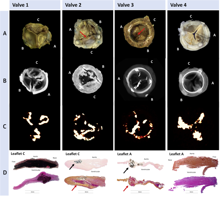
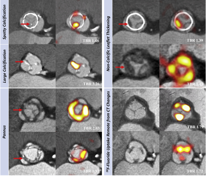
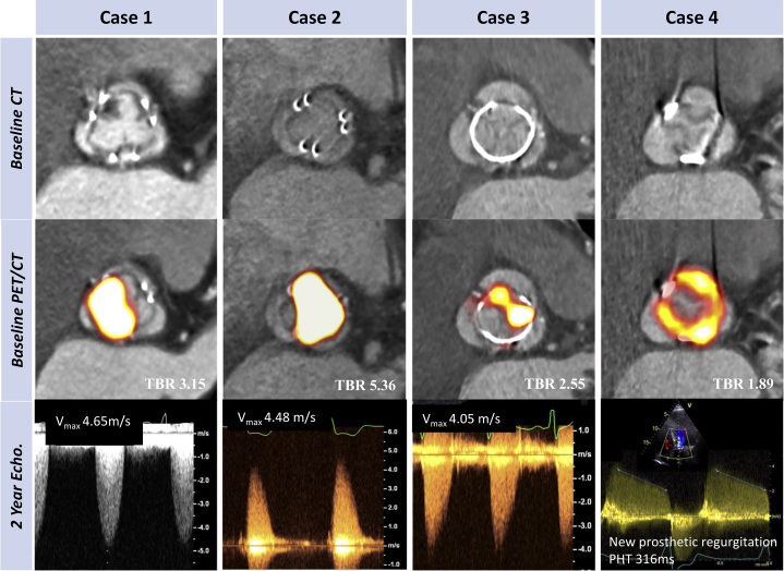
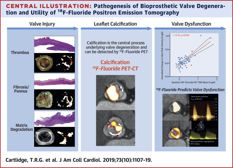
Results: All ex vivo, degenerate bioprosthetic valves displayed 18F-fluoride PET uptake that colocalized with tissue degeneration on histology. In 71 patients without known bioprosthesis dysfunction, 14 had abnormal leaflet pathology on CT, and 24 demonstrated 18F-fluoride PET uptake (target-to-background ratio 1.55 [interquartile range (IQR): 1.44 to 1.88]). Patients with increased 18F-fluoride uptake exhibited more rapid deterioration in valve function compared with those without (annualized change in peak transvalvular velocity 0.30 [IQR: 0.13 to 0.61] vs. 0.01 [IQR: -0.05 to 0.16] ms-1/year; p < 0.001). Indeed 18F-fluoride uptake correlated with deterioration in all the conventional echocardiographic measures of valve function assessed (e.g., change in peak velocity, r = 0.72; p < 0.001). Each of the 10 patients who developed new overt bioprosthesis dysfunction during follow-up had evidence of 18F-fluoride uptake at baseline (target-to-background ratio 1.89 [IQR: 1.46 to 2.59]). On multivariable analysis, 18F-fluoride uptake was the only independent predictor of future bioprosthetic dysfunction.

Conclusions: 18F-fluoride PET-CT identifies subclinical bioprosthetic valve degeneration, providing powerful prediction of subsequent valvular dysfunction and highlighting patients at risk of valve failure. This technique holds major promise in the diagnosis of valvular degeneration and the surveillance of patients with bioprosthetic valves. (18F-Fluoride Assessment of Aortic Bioprosthesis Durability and Outcome [18F-FAABULOUS]; NCT02304276).

Keywords: aortic valve replacement; bioprosthetic valve degeneration; calcification; histology; positron emission tomography.


Figures

None
Graphical abstract
Figure 1
Figure 1
Ex Vivo Degenerated Bioprosthetic Aortic Valves: Macroscopic Appearances, Micro-CT, Micro-PET, and Histology (Row A) Macroscopic visual appearances of failed and explanted bioprosthetic valves. (Row B) CT en face images of the valves. (Row C) PET en face images demonstrating increased 18F-fluoride uptake in all valves. (Row D) Histology staining of sections taken from valve leaflet as indicated, with von Kossa (top row, calcium appears black), Movat Pentachrome (bottom row, valves 1 and 4), and hematoxylin and eosin (bottom row, valves 2 and 3) stains. All 4 degenerate bioprostheses demonstrate increased 18F-fluoride uptake in the valve leaflets. In valve 1, this uptake corresponds to gross leaflet calcification observed macroscopically and on CT images with confirmation on histology (extensive black staining). In valve 2, increased 18F-fluoride uptake is observed in association with fibrotic leaflet thickening and pannus (red arrows) with associated calcification (black arrows) observed macroscopically and on CT with confirmation on histology. In valve 3, increased 18F-fluoride uptake is observed at the site of valve leaflet thrombus (red arrow) observed macroscopically at the base of leaflet 1, with confirmation of thrombus (red arrow) and colocalized calcification (black arrow) on histology. In valve 4, extensive 18F-fluoride uptake is observed in the absence of calcification on CT and histology but instead in areas of leaflet thickening, marked fluid insudation, and disrupted collagen architecture. CT = computed tomography; PET = positron emission tomography.
Figure 2
Figure 2
In Vivo 18F-fluoride PET and CT Imaging of Patients With Bioprosthetic Aortic Valves Baseline CT (left) and 18F-fluoride PET (right) images from patients with bioprosthetic aortic valves. En face CT images of aortic bioprosthetic valves showing spotty calcification and large calcification (top left), circumferential pannus (bottom left), and noncalcific leaflet thickening suggestive of thrombus (top right) (all abnormalities identified by red arrows). Hybrid en face PET-CT images in the same patients: increased bioprosthetic 18F-fluoride activity (red/yellow areas) is observed in each patient colocalizing with the CT abnormalities. 18F-fluoride activity was also commonly observed remote from leaflet changes on CT (bottom right). Target-to-background (TBR) values are annotated on the hybrid PET-CT images in white text. Abbreviations as in Figure 1.
Figure 3
Figure 3
Baseline 18F-fluoride PET Uptake Predicts Subsequent Deterioration in Bioprosthetic Valve Function After 2 Years (A) A strong correlation was observed between baseline 18F-fluoride uptake in the bioprosthetic valves (TBR) and subsequent progression in bioprosthetic valve peak velocity (log transformation applied; r = 0.72; p < 0.001). Orange dots signify patients who developed new bioprosthetic valve regurgitation during follow-up. (B) 18F-fluoride uptake (dashed orange line represents threshold for increased 18F-fluoride uptake; TBR 1.3) in patients with different stages of structural valve degeneration after 2-year follow-up (stage 0: no significant change from post-implantation [n = 54]; stage 1: morphological abnormalities without significant hemodynamic changes [n = 9]; stage 2: new moderate stenosis and/or regurgitation [n = 5]; stage 3: new severe stenosis and/or severe regurgitation [n = 2]) demonstrating incrementally higher uptake values with increasing severity of structural valve degeneration. (C and D) Forest plots of unstandardized coefficients (95% confidence intervals) from a multivariable linear regression analysis predicting change in bioprosthetic valve function (annualized change in peak velocity) during follow-up. When examining all relevant baseline characteristics, 18F-fluoride uptake was the only independent predictor of hemodynamic deterioration in valve function when used both as a dichotomous variable (PET+, TBR >1.3) (C) and as a continuous variable (TBR) (D). CI = confidence interval; other abbreviations as in Figures 1 and 2.
Figure 4
Figure 4
Case Illustrations: Baseline 18F-fluoride PET and CT Predict Imminent Failure of Bioprosthetic Function Cases 1 to 4 illustrate the utility of 18F-fluoride PET and CT in the prediction of deteriorating valve performance. None of the patients had known bioprosthetic degeneration at baseline. En face contrast-enhanced CT images (top row) demonstrate noncalcific leaflet thickening in case 1, but no clear structural CT changes in the remaining cases. Hybrid 18F-fluoride PET-CT images (middle row) demonstrate high-intensity 18F-fluoride activity in all the valves (TBR values in white). Doppler echocardiographic assessments of bioprosthetic valve function after follow-up (bottom row). In each case, new valve dysfunction has developed with progression to severe obstruction in cases 1, 2, and 3 and new moderate/severe eccentric regurgitation in case 4 (pressure half time 316 ms with holodiastolic flow reversal in aorta). Patient #1 died from valve-related heart failure, Patient #2 required redo surgical valve replacement, Patient #3 remains under close surveillance, and Patient #4 is undergoing work-up for redo surgery.
Central Illustration
Pathogenesis of Bioprosthetic Valve Degeneration and Utility of 18F-Fluoride Positron Emission Tomography (Left column) Potential mechanisms of initial valve injury include leaflet thrombosis, fibrosis, and matrix degradation with expansion of the central proteoglycan layer, fluid insudation, and disruption of normal collagen architecture. (Middle column) Each appears to lead to bioprosthetic valve calcification as the final common pathway for valve degeneration that can be detected with 18F-fluoride PET imaging. (Right column) Baseline 18F-fluoride uptake correlates strongly with subsequent deterioration in valve function during follow-up (annualized change in peak transvalvular velocity: r = 0.72; p < 0.001) (top). Two examples demonstrate high-intensity 18F-fluoride leaflet uptake at baseline in valves without previous evidence of degeneration. After follow-up, both bioprosthetic valves developed clear evidence of dysfunction: one with severe stenosis, and the other, regurgitation. PET = positron emission tomography.

Comment in

References

    1. Iung B., Vahanian A. Epidemiology of valvular heart disease in the adult. Nat Rev Cardiol. 2011;8:162–172. - PubMed
    1. Nkomo V.T., Gardin J.M., Skelton T.N., Gottdiener J.S., Scott C.G., Enriquez-Sarano M. Burden of valvular heart diseases: a population-based study. Lancet. 2006;368:1005–1011. - PubMed
    1. Brown J.M., O’Brien S.M., Wu C., Sikora J.A., Griffith B.P., Gammie J.S. Isolated aortic valve replacement in North America comprising 108,687 patients in 10 years: changes in risks, valve types, and outcomes in the Society of Thoracic Surgeons National Database. J Thorac Cardiov Sur. 2009;137:82–90. - PubMed
    1. Brennan J.M., Thomas L., Cohen D. Transcatheter versus surgical aortic valve replacement: propensity-matched comparison. J Am Coll Cardiol. 2017;70:439–450. - PMC - PubMed
    1. Pibarot P., Dumesnil J.G. Prosthetic heart valves: selection of the optimal prosthesis and long-term management. Circulation. 2009;119:1034–1048. - PubMed

Publication types

MeSH terms

Substances

Associated data