Review
. 2019 Mar 19;73(10):1189-1206.
doi: 10.1016/j.jacc.2018.12.064.

Autonomic Nervous System Dysfunction: JACC Focus Seminar

Affiliations
Review

Autonomic Nervous System Dysfunction: JACC Focus Seminar

Jeffrey J Goldberger et al. J Am Coll Cardiol. .

Abstract

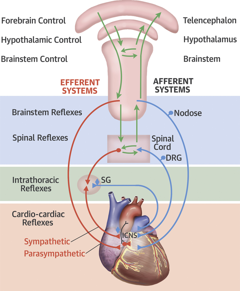
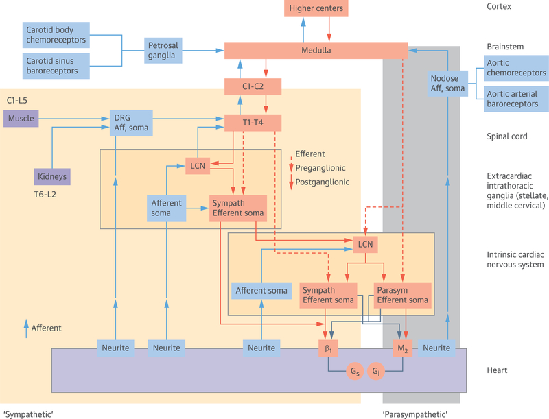
Autonomic nervous system control of the heart is a dynamic process in both health and disease. A multilevel neural network is responsible for control of chronotropy, lusitropy, dromotropy, and inotropy. Intrinsic autonomic dysfunction arises from diseases that directly affect the autonomic nerves, such as diabetes mellitus and the syndromes of primary autonomic failure. Extrinsic autonomic dysfunction reflects the changes in autonomic function that are secondarily induced by cardiac or other disease. An array of tests interrogate various aspects of cardiac autonomic control in either resting conditions or with physiological perturbations from resting conditions. The prognostic significance of these assessments have been well established. Clinical usefulness has not been established, and the precise mechanistic link to mortality is less well established. Further efforts are required to develop optimal approaches to delineate cardiac autonomic dysfunction and its adverse effects to develop tools that can be used to guide clinical decision-making.

Keywords: arrhythmia; autonomic; heart failure; myocardial infarction.


Figures

FIGURE 1
FIGURE 1. Autonomic Neural Control of the Heart
Autonomic neural control of the heart. Modified with permission from Shivkumar et al. (4). DRG= dorsal root ganglion; ICNS = intrinsic nervous system; SG = stellate ganglion.
FIGURE 2
FIGURE 2. Functional Organization of Cardiac Neural Control
The shaded areas (yellow = sympathetic fibers, grey = parasympathetic fibers [vagal trunk]) roughly correspond to the anatomical sympathetic and parasympathetic nerve fibers that are seen macroscopically. The afferent neural fibers run along with nerves anatomically referred to as sympathetic and parasympathetic trunks. Modified with permission from Shivkumar et al. (4). Aff = afferent; LCN = local circuit neuron; other abbreviation as Figure 1.
FIGURE 3
FIGURE 3. Diagram of the QT-RR Relationship
The QT-RR relationship in the early post-exercise recovery period without autonomic blockade (baseline [orange]), with beta-blockade (green), with parasympathetic blockade (atropine [grey]), and double blockade with propranolol and atropine (blue). Original data from Sundaram et al. (160).

References

    1. Vaseghi M, Barwad P, Malavassi Corrales FJ, et al. Cardiac sympathetic denervation for refractory ventricular arrhythmias. J Am Coll Cardiol 2017;69:3070–80. - PMC - PubMed
    1. Armour JA, Murphy DA, Yuan BX, Macdonald S, Hopkins DA. Gross and microscopic anatomy of the human intrinsic cardiac nervous system. Anat Rec 1997;247:289–98. - PubMed
    1. Buckley U, Shivkumar K, Ardell JL. Autonomic regulation therapy in heart failure. Curr Heart Fail Rep 2015;12:284–93. - PMC - PubMed
    1. Shivkumar K, Ajijola OA, Anand I, et al. Clinical neurocardiology defining the value of neuroscience-based cardiovascular therapeutics. J Physiol 2016;594:3911–54. - PMC - PubMed
    1. Armour JA. Cardiac neuronal hierarchy in health and disease. Am J Physiol Regul Integr Comp Physiol 2004;287:R262–71. - PubMed

Publication types

MeSH terms