

Repeatability and reproducibility of longitudinal relaxation rate in 12 small-animal MRI systems
- PMID: 30872166
- PMCID: PMC6477178
- DOI: 10.1016/j.mri.2019.03.008
Repeatability and reproducibility of longitudinal relaxation rate in 12 small-animal MRI systems
Abstract
Background: Many translational MR biomarkers derive from measurements of the water proton longitudinal relaxation rate R1, but evidence for between-site reproducibility of R1 in small-animal MRI is lacking.
Objective: To assess R1 repeatability and multi-site reproducibility in phantoms for preclinical MRI.
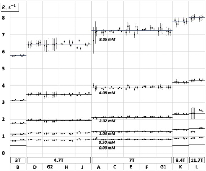
Methods: R1 was measured by saturation recovery in 2% agarose phantoms with five nickel chloride concentrations in 12 magnets at 5 field strengths in 11 centres on two different occasions within 1-13 days. R1 was analysed in three different regions of interest, giving 360 measurements in total. Root-mean-square repeatability and reproducibility coefficients of variation (CoV) were calculated. Propagation of reproducibility errors into 21 translational MR measurements and biomarkers was estimated. Relaxivities were calculated. Dynamic signal stability was also measured.
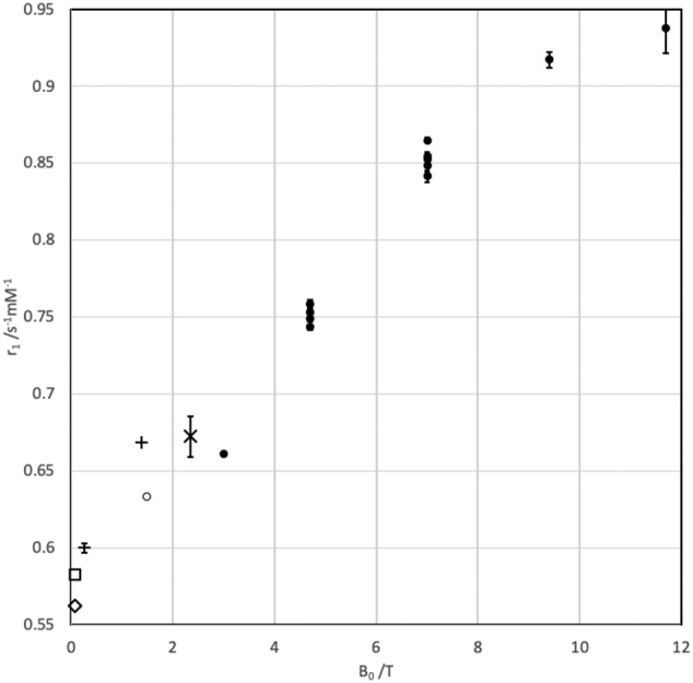
Results: CoV for day-to-day repeatability (N = 180 regions of interest) was 2.34% and for between-centre reproducibility (N = 9 centres) was 1.43%. Mostly, these do not propagate to biologically significant between-centre error, although a few R1-based MR biomarkers were found to be quite sensitive even to such small errors in R1, notably in myocardial fibrosis, in white matter, and in oxygen-enhanced MRI. The relaxivity of aqueous Ni2+ in 2% agarose varied between 0.66 s-1 mM-1 at 3 T and 0.94 s-1 mM-1 at 11.7T.
Interpretation: While several factors affect the reproducibility of R1-based MR biomarkers measured preclinically, between-centre propagation of errors arising from intrinsic equipment irreproducibility should in most cases be small. However, in a few specific cases exceptional efforts might be required to ensure R1-reproducibility.
Keywords: Biomarker; Error propagation; Hardware stability; MRI; Phantom; Relaxation time; Reproducibility.
Copyright © 2019 The Authors. Published by Elsevier Inc. All rights reserved.
Figures
References
-
- Stikov N., Boudreau M., Levesque I.R., Tardif C.L., Barral J.K., Pike G.B. On the accuracy of T1 mapping: searching for common ground. Magn Reson Med. 2015;73:514–522. - PubMed
-
- Keenan K.E., Ainslie M., Barker A.J., Boss M.A., Cecil K.M., Charles C. Quantitative magnetic resonance imaging phantoms: a review and the need for a system phantom. Magn Reson Med. 2018;79:48–61. - PubMed
-
- Captur G., Gatehouse P., Keenan K.E., Heslinga F.G., Bruehl R., Prothmann M. A medical device-grade T1 and ECV phantom for global T1 mapping quality assurance - the T1 mapping and ECV standardization in cardiovascular magnetic resonance (T1MES) program. J Cardiovasc Magn Reson. 2016;18:1–20. - PMC - PubMed
-
- Vassiliou V.S., Heng E.L., Gatehouse P.D., Donovan J., Raphael C.E., Giri S. Magnetic resonance imaging phantoms for quality-control of myocardial T1 and ECV mapping: specific formulation, long-term stability and variation with heart rate and temperature. J Cardiovasc Magn Reson. 2016;18:1–12. - PMC - PubMed
-
- Lerski R.A., McRobbie D.W., Straughan K., Walker P.M., de Certaines J.D., Bernard A.M. V. Multi-center trial with protocols and prototype test objects for the assessment of MRI equipment. Magn Reson Imaging. 1988;6:201–214. - PubMed
Publication types
MeSH terms
Substances
Grants and funding
LinkOut - more resources
Full Text Sources
Medical
