Review
. 2019 Mar;19(2):169-176.
doi: 10.7861/clinmedicine.19-2-169.

Neuromyelitis optica spectrum disorders

Affiliations
Review

Neuromyelitis optica spectrum disorders

Saif Huda et al. Clin Med (Lond). 2019 Mar.

Abstract

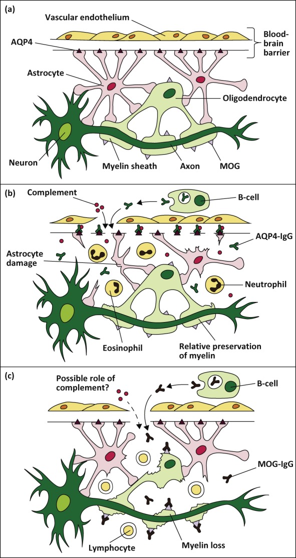
Neuromyelitis optica spectrum disorder (NMOSD) is an uncommon antibody-mediated disease of the central nervous system. Long segments of spinal cord inflammation (myelitis), severe optic neuritis, and/or bouts of intractable vomiting and hiccoughs (area postrema syndrome) are classic presentations of the disease and may alert the clinician to the diagnosis. Untreated, approximately 50% of NMOSD patients will be wheelchair users and blind, and a third will have died within 5 years of their first attack. Unlike multiple sclerosis, a progressive clinical course is very unusual and the accrual of disability is related to relapses. Approximately 75% of patients have antibodies against aquaporin-4, a water channel expressed on astrocytes. Relapses are treated aggressively to prevent residual disability with high-dose steroids and often plasma exchange. Relapse prevention is crucial and achieved with long-term immunosuppression. In this article we review the pathogenesis, clinical features, diagnosis and management of NMOSD.

Keywords: Neuromyelitis optica; antibody; aquaporin-4.


Figures

Fig 1.
Fig 1.
(a) This figure illustrates the sites of expression of aquaporin-4 (AQP4) and myelin oligodendrocyte glycoprotein (MOG) in the central nervous system (CNS). AQP4 is expressed on astrocyte 'foot-like' processes at the blood-brain barrier. MOG is expressed on oligodendrocytes on the outermost lamellae of myelin sheaths. (b) AQP4-Abs (IgG) are produced systemically by mature B-cells, and upon crossing the blood-brain barrier, activate complement-mediated astrocyte damage. There is relative preservation of myelin initially. The inflammatory milieu consists of neutrophils and eosiniphils. (c) MOG-Abs (IgG) are also produced outside the CNS, and causes demyelination by mechanisms that are poorly understood. Reprinted with permission from Whittam D, Wilson M, Hamid S et al. What’s new in neuromyelitis optica? A short review for the clinical neurologist. J Neurol 2017;264:2330–44.
Fig 2.
Fig 2.
Magnetic resonance imaging (MRI) in neuromyelitis optica. (a) Longitudinally extensive transverse myelitis – sagittal T2 weighted image of cervicothoracic spine demonstrating T2 hyperintensity extending longitudinally from C2–T4. (b) Area postrema syndrome – axial T2 weighted image demonstrating dorsal T2 signal hyperintensity in the medulla. (c) Brainstem syndrome – axial T2 weighted image demonstrating diffuse symmetrical T2 signal hyperintensity in the medulla.

References

    1. Jarius S, Wildemann B. The history of neuromyelitis optica. J Neuroinflammation 2013;10:8. - PMC - PubMed
    1. Lennon VA, Wingerchuk DM, Kryzer TJ, et al. A serum autoantibody marker of neuromyelitis optica: distinction from multiple sclerosis. Lancet 2004;364:2106–12. - PubMed
    1. Wingerchuk DM, Banwell B, Bennett JL, et al. International consensus diagnostic criteria for neuromyelitis optica spectrum disorders. Neurology 2015; 85:177–89. - PMC - PubMed
    1. Kitley J, Woodhall M, Waters P, et al. Myelin-oligodendrocyte glycoprotein antibodies in adults with a neuromyelitis optica phenotype. Neurology 2012;79:1273–7. - PubMed
    1. Lennon VA, Kryzer TJ, Pittock SJ, Verkman AS, Hinson SR. IgG marker of optic-spinal multiple sclerosis binds to the aquaporin-4 water channel. J Exp Med 2005;202:473–7. - PMC - PubMed