. 2019 Dec;68(12):2186-2194.
doi: 10.1136/gutjnl-2018-317458. Epub 2019 Mar 14.

Enrichment of oral microbiota in early cystic precursors to invasive pancreatic cancer

Affiliations

Enrichment of oral microbiota in early cystic precursors to invasive pancreatic cancer

Rogier Aäron Gaiser et al. Gut. 2019 Dec.

Abstract

Objectives: Intraductal papillary mucinous neoplasms (IPMNs) are pancreatic cysts that can progress to invasive pancreatic cancer. Associations between oncogenesis and oral microbiome alterations have been reported. This study aims to investigate a potential intracystic pancreatic microbiome in a pancreatic cystic neoplasm (PCN) surgery patient cohort.

Design: Paired cyst fluid and plasma were collected at pancreatic surgery from patients with suspected PCN (n=105). Quantitative and qualitative assessment of bacterial DNA by qPCR, PacBio sequencing (n=35), and interleukin (IL)-1β quantification was performed. The data were correlated to diagnosis, lesion severity and clinical and laboratory profile, including proton-pump inhibitor (PPI) usage and history of invasive endoscopy procedures.

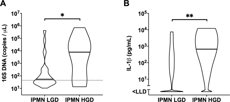
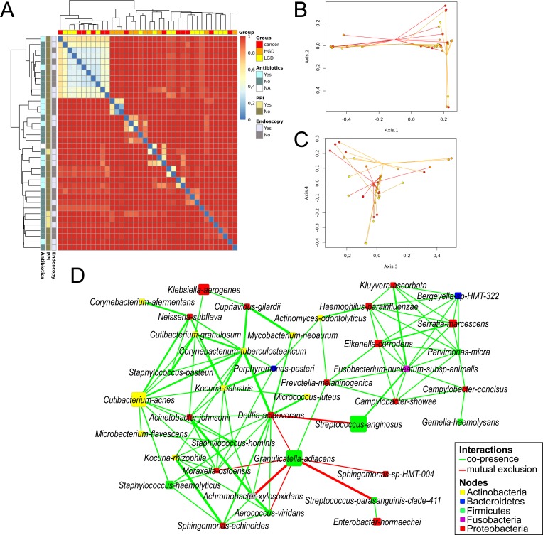
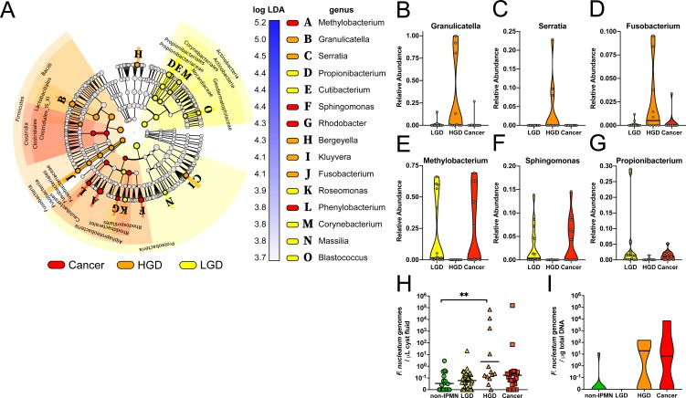
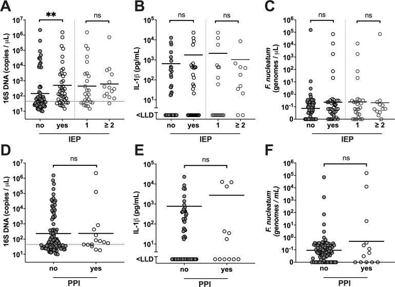
Results: Intracystic bacterial 16S DNA copy number and IL-1β protein quantity were significantly higher in IPMN with high-grade dysplasia and IPMN with cancer compared with non-IPMN PCNs. Despite high interpersonal variation of intracystic microbiota composition, bacterial network and linear discriminant analysis effect size analyses demonstrated co-occurrence and enrichment of oral bacterial taxa including Fusobacterium nucleatum and Granulicatella adiacens in cyst fluid from IPMN with high-grade dysplasia. The elevated intracystic bacterial DNA is associated with, but not limited to, prior exposure to invasive endoscopic procedures, and is independent from use of PPI and antibiotics.

Conclusions: Collectively, these findings warrant further investigation into the role of oral bacteria in cystic precursors to pancreatic cancer and have added values on the aetiopathology as well as the management of pancreatic cysts.

Keywords: bacterial translocation; endoscopic procedure; inflammation; pancreatic surgery; pancreatic tumours.


Conflict of interest statement

Competing interests: None declared.

Figures

Figure 1
Figure 1
Intracystic quantities of (A) bacterial 16S DNA and (B) IL-1β in ’non-IPMN' (n=21), ’IPMN' (n=57), ’IPMN Cancer' (n=27) patients, and in paired plasma (D, E) of ’non-IPMN' (n=10), ’IPMN' (n=15), ’IPMN Cancer' (n=7) patients. Dotted line indicates assay cut-off value. Geometric mean for 16S DNA and mean for IL-1β are indicated per each group. Spearman’s correlation analysis on 16S DNA and IL-1β quantities in (C) cyst fluid and (F) plasma, displaying non-linear fit curve (solid line) and 95% CI (dashed line). Statistically significant differences were determined using Kruskal-Wallis test with Dunn’s multiple comparison test with *p<0.05, **p<0.01, ***p<0.001. IL, interleukin; IPMN, intraductal papillary mucinous neoplasm; LLD, lower limit of detection; n.d., not determined.
Figure 2
Figure 2
Violin plot showing the distribution with median of (A) total bacterial 16S DNA in ’IPMN LGD' (n=45) and ’IPMN HGD' (n=14) and (B) IL-1β in ’IPMN LGD' (n=35) and ’IPMN HGD' (n=10) cyst fluid samples, determined by TaqMan qPCR and ELISA, respectively. Statistical comparisons between groups were made using Mann-Whitney U test with *p<0.05 and **p<0.01. Dotted line indicates assay cut-off value. HGD, high-grade dysplasia; IL, interleukin; IPMN, intraductal papillary mucinous neoplasm; LGD, low-grade dysplasia; LLD, lower limit of detection.
Figure 3
Figure 3
Box plot displaying (A) Chao1 richness (white), (B) Shannon entropy (light grey) and (C) inverted Simpson’s (dark grey) indices per diagnose group at operational taxonomic unit (OTU) level. Bar plots showing relative abundance of OTUs per sample at (D) phylum level and (E) genus level. HGD, high-grade dysplasia; IPMN, intraductal papillary mucinous neoplasm; LGD, low-grade dysplasia.
Figure 4
Figure 4
Beta diversity between samples visualised by (A) clustered heatmap showing Bray-Curtis dissimilarity at OTU level for each sample, characterised horizontally by diagnose group and vertically by previous endoscopic procedures, PPI use and antibiotic medication <1 month prior to surgery. (B) PCoA spider plots displaying dimensions 1 and 2 and (C) dimensions 3 and 4. Samples are coloured according to final diagnose (yellow=IPMN LGD, orange=IPMN HGD, red=IPMN Cancer). (D) Bacterial co-occurrence and antioccurrence visualised in a network, with nodes representing bacterial species (coloured according to phylum, size corresponds to mean relative abundance) and edges representing interactions (green=co-occurrence, red=mutual exclusion) supported by >3 methods at statistical significance of q<0.01. HGD, high-grade dysplasia; IPMN, intraductal papillary mucinous neoplasm; LGD, low-grade dysplasia; PPI, proton-pump inhibitor.
Figure 5
Figure 5
(A) Cladogram representing linear discriminant analysis effect size results which identified 15 features that are differentially abundant between the three diagnose classes (red=Cancer, orange=IPMN HGD, yellow=IPMN LGD). (B–G) Violin plot showing relative abundance distribution and median of selected differentially abundant bacterial genera per diagnose group. Validation of Fusobacterium nucleatum genome quantification by TaqMan qPCR in (H) cyst fluid and (I) DNA extracted from 25 formalin-fixed PCN tissue samples (non-IPMN n=9, IPMN LGD n=6, IPMN HGD n=4, Cancer n=6). Statistically significant differences were determined using Kruskal-Wallis test with Dunn’s multiple comparison test **p<0.01. HGD, high-grade dysplasia; IPMN, intraductal papillary mucinous neoplasm; LGD, low-grade dysplasia; PCN, pancreatic cystic neoplasm.
Figure 6
Figure 6
Patient stratification based on previous pancreatic invasive procedures (IEP), classified as ’no' (n=65) and ’yes' (n=40), comprising those with '1' IEP (n=26) and '≥2' (n=14), or use of proton-pump inhibitors (PPI), classified as ’no' (n=90) and ’yes' (n=15). Panels display intracystic (A) 16S DNA quantities, (B) interleukin (IL)-1β quantity and (C) Fusobacterium nucleatum genome copies, with the geometric mean value indicated per group stratified on IEP, and intracystic (D) 16S DNA quantities, (E) IL-1β quantity and (F) F. nucleatum genome copies, with the geometric mean value indicated per group stratified on PPI use. Statistically significant differences between ’no' and ’yes' groups, and '1' and '≥2' groups, respectively, were determined using Mann-Whitney U test, with **p<0.01. LLD, lower limit of detection; ns, not significant.

Comment in

References

    1. European Study Group on Cystic Tumours of the Pancreas. European evidence-based guidelines on pancreatic cystic neoplasms. Gut 2018;67:789–804. 10.1136/gutjnl-2018-316027 - DOI - PMC - PubMed
    1. Chandwani R, Allen PJ. Cystic Neoplasms of the Pancreas. Annu Rev Med 2016;67:45–57. 10.1146/annurev-med-051914-022011 - DOI - PubMed
    1. Valsangkar NP, Morales-Oyarvide V, Thayer SP, et al. 851 resected cystic tumors of the pancreas: a 33-year experience at the Massachusetts General Hospital. Surgery 2012;152:S4–12. 10.1016/j.surg.2012.05.033 - DOI - PMC - PubMed
    1. Ferrone CR, Correa-Gallego C, Warshaw AL, et al. Current trends in pancreatic cystic neoplasms. Arch Surg 2009;144:448–54. 10.1001/archsurg.2009.36 - DOI - PMC - PubMed
    1. Aronsson L, Andersson R, Ansari D. Intraductal papillary mucinous neoplasm of the pancreas - epidemiology, risk factors, diagnosis, and management. Scand J Gastroenterol 2017;52:803–15. 10.1080/00365521.2017.1318948 - DOI - PubMed

Publication types

MeSH terms