. 2019 May 10;294(19):7588-7600.
doi: 10.1074/jbc.RA118.006233. Epub 2019 Mar 14.

Residues in the fingers domain of the translesion DNA polymerase DinB enable its unique participation in error-prone double-strand break repair

Affiliations

Residues in the fingers domain of the translesion DNA polymerase DinB enable its unique participation in error-prone double-strand break repair

Tommy F Tashjian et al. J Biol Chem. .

Abstract

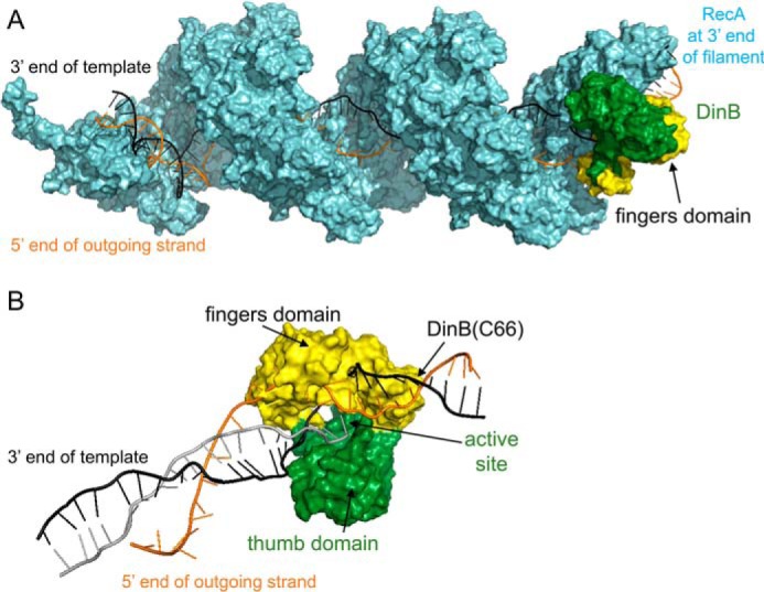
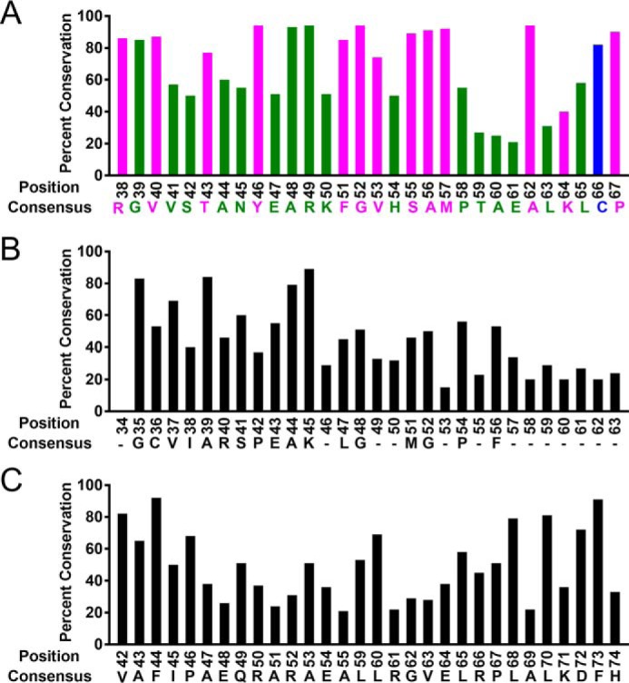
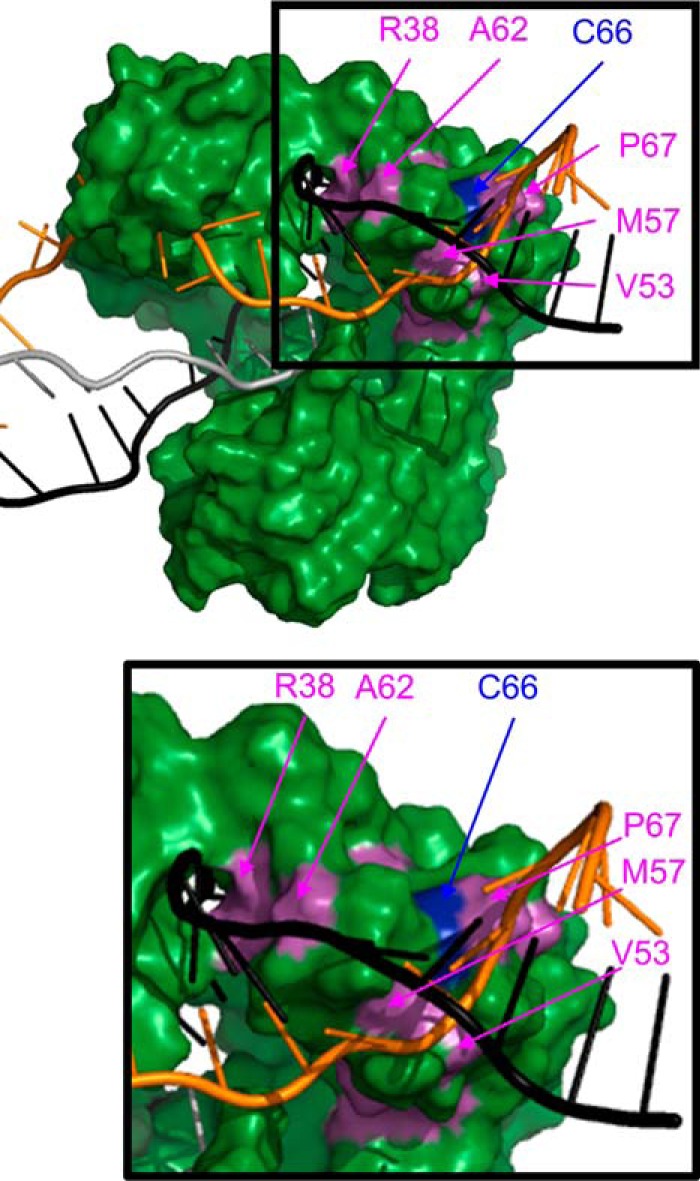
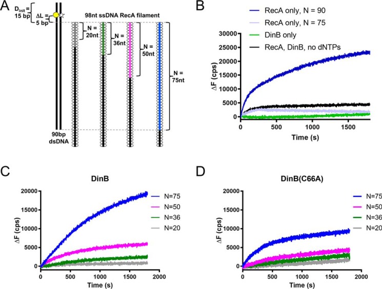
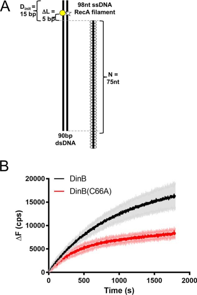
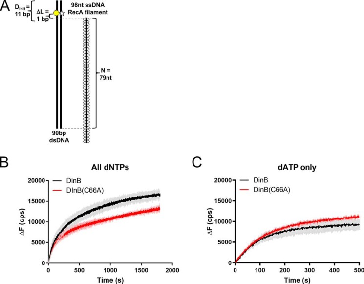
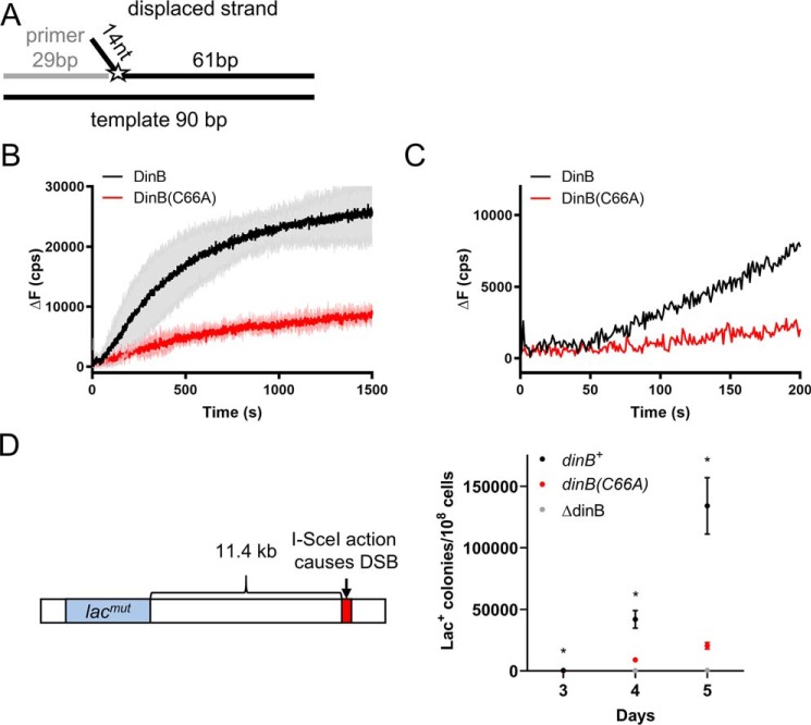
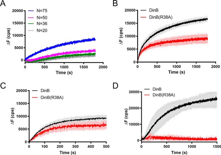
The evolutionarily conserved Escherichia coli translesion DNA polymerase IV (DinB) is one of three enzymes that can bypass potentially deadly DNA lesions on the template strand during DNA replication. Remarkably, however, DinB is the only known translesion DNA polymerase active in RecA-mediated strand exchange during error-prone double-strand break repair. In this process, a single-stranded DNA (ssDNA)-RecA nucleoprotein filament invades homologous dsDNA, pairing the ssDNA with the complementary strand in the dsDNA. When exchange reaches the 3' end of the ssDNA, a DNA polymerase can add nucleotides onto the end, using one strand of dsDNA as a template and displacing the other. It is unknown what makes DinB uniquely capable of participating in this reaction. To explore this topic, we performed molecular modeling of DinB's interactions with the RecA filament during strand exchange, identifying key contacts made with residues in the DinB fingers domain. These residues are highly conserved in DinB, but not in other translesion DNA polymerases. Using a novel FRET-based assay, we found that DinB variants with mutations in these conserved residues are less effective at stabilizing RecA-mediated strand exchange than native DinB. Furthermore, these variants are specifically deficient in strand displacement in the absence of RecA filament. We propose that the amino acid patch of highly conserved residues in DinB-like proteins provides a mechanistic explanation for DinB's function in strand exchange and improves our understanding of recombination by providing evidence that RecA plays a role in facilitating DinB's activity during strand exchange.

Keywords: DNA damage; DNA polymerase; DNA polymerase IV; DNA repair; DNA synthesis; DinB; RecA; homologous recombination.


Conflict of interest statement

The authors declare that they have no conflicts of interest with the contents of this article

Figures

Figure 1.
Figure 1.
Molecular modeling of DinB in RecA-mediated strand exchange indicates the DinB fingers domain separates dsDNA. A, this model of RecA–DNA–DinB interaction represents the homologous recombination stage where strand exchange has taken place up to the 3′ extremity of the incoming strand. Interactions were modeled using structures and models as follows. The ssDNA (gray, largely obscured by RecA), template strand of dsDNA (black), displaced strand of dsDNA (orange), and RecA (cyan) were modeled using the PDB structure 3CMW (51) that had been extended by three turns (52) in a previous model (53). DinB (green and yellow) was modeled using the PDB structure 4IRC (43). The RecA nucleoprotein filament and DinB are shown interacting with the RecA molecule that lies at the 3′ end of the nucleoprotein filament. DinB and the RecA nucleoprotein filament were surface-rendered with PyMOL (42). The fingers domain of DinB is highlighted in yellow. B, DinB and DNA are shown without RecA to permit visualization of DinB's interaction with three DNA strands during strand exchange. Structures have been turned ∼180° about the horizontal axis with respect to A. DinB's fingers domains (yellow) appears to separate the template (black) and displaced (orange) strands of the dsDNA.
Figure 2.
Figure 2.
Several residues of the DinB fingers domain are highly conserved only in DinB-like proteins. Multiple sequence alignments show that an amino acid patch, depicted here in magenta and blue (A) located in the fingers domain of DinB, is highly conserved in DinB-like proteins but not in DNA polymerase V (B) or in DNA polymerase II (C) homologs. Percent conservation values for A–C are reported in Table S1.
Figure 3.
Figure 3.
Modeling suggests that several conserved DinB residues are located near the template (black) and displaced (orange) strands during strand exchange. Depiction of DinB from surface-rendered model from Fig. 1 shows that cysteine 66 (blue) and other predominantly conserved residues only in DinB-like proteins (magenta) are located near the template (Arg-38 and Ala-62) and displaced (Val-53, Met-57, Cys-66, and Pro-67) strands of dsDNA during strand exchange. Enlargement of the boxed area is provided to better visualize the positions of the highlighted residues. Residues in green, magenta, and blue color correspond to residues of the same color in Fig. 2.
Figure 4.
Figure 4.
Highly conserved fingers domain residue is important for DinB's activity during strand exchange. A, schematic shows experimental setup for RecA-dependent strand-exchange experiments. Fluorescently-labeled dsDNA (left) is mixed with each of a set of ssDNA RecA filaments (right). Each in the set of filaments contains a different length of homology (N) to the labeled dsDNA (signified by gray, green, magenta, and blue lines). In the dsDNA, the template strand is labeled with rhodamine (yellow circle), and the displaced strand is labeled with fluorescein (white star). These fluorophores are located 5 bp away from area of homology between dsDNA and ssDNA RecA filament (ΔL = 5 bp). The proximity of the rhodamine quenches fluorescein fluorescence until template and displaced strands are separated. Fluorescence increases when the ssDNA RecA filament invades the dsDNA, and DinB synthesizes DNA using the ssDNA filament as a primer. Five nucleotide insertions are needed to separate the displaced strand at the location of the fluorescent labels and relieve quenching. B, when only RecA or only DinB is mixed with the highest homology ssDNA RecA filament (N = 75, blue filament in A), the fluorescent labels are not efficiently separated, indicating that the dsDNA is still annealed at the location of the labels. When both proteins are present in the absence of dCTP, dGTP, and dTTP (dATP is present for nucleoprotein filament assembly), baseline fluorescence is observed. A RecA filament with full homology to the dsDNA (N = 90) is used to determine maximum possible fluorescence in the assay. C, DinB stabilizes strand exchange in a homology-dependent manner. As homology increases between the ssDNA RecA filament and the fluorescently-labeled dsDNA, DinB efficiently separates the dsDNA at the location of the fluorescent labels. This indicates that increased homology allows DinB to more efficiently stabilize strand-exchange products. D, DinB(C66A) stabilizes strand-exchange products, but it does so with less efficiency than the native enzyme. N indicates the length of homology between dsDNA and ssDNA filament; ΔL indicates the distance between region of homology on dsDNA and fluorescent label; Dinit indicates the distance between fluorescent label and closet end of dsDNA; ΔF indicates the change in fluorescence measured in counts/s with respect to the fluorescence at 0 s. Experiments were performed in triplicate with similar results. Representative data are shown.
Figure 5.
Figure 5.
DinB stabilizes strand-exchange products significantly better than DinB(C66A). A, as in Fig. 4, RecA-dependent strand-exchange experiments utilize an ssDNA RecA filament and dsDNA that is fluorescently labeled on either strand to cause quenching. These fluorophores are located 5 bp away from the region of homology between the dsDNA and the ssDNA RecA filament (ΔL = 5 bp). Shown here is the ssDNA RecA filament with the highest homology, 75 nt. Experiments were performed using all four dNTPs. B, shown is the direct comparison between the fluorescence obtained with DinB and DinB(C66A) in experiments with 75 bp of homology between the RecA ssDNA filament and the dsDNA (directly compares blue lines from Fig. 4, C and D). Both DinB and DinB(C66A) stabilize strand-exchange products and separate the dsDNA at the location of the labels, but DinB does this better than the variant. The DinB reaction has a significantly higher initial rate (18.59 ± 0.3395 cps) than the DinB(C66A) reaction (14.34 ± 0.2784 cps, p value < 0.0001). The DinB reaction also reaches a significantly higher maximum ΔF (16,987 ± 2519 cps) than the DinB(C66A) reaction (8893 ± 913; p value < 0.01). N indicates the length of homology between dsDNA and ssDNA filament; ΔL indicates the distance between region of homology on dsDNA and fluorescent label; Dinit indicates the distance between fluorescent label and closet end of dsDNA; ΔF indicates the change in fluorescence measured in counts/s with respect to the fluorescence at 0 s. Experiments were performed in triplicate. Mean ± S.D. (shaded region surrounding curves) is shown.
Figure 6.
Figure 6.
DinB(C66A) can insert a single nucleotide during strand exchange as well as DinB. A, RecA-dependent strand-exchange experiments were performed similarly to Fig. 5, except fluorophores are located 1 bp away from the region of homology between the dsDNA and the ssDNA RecA filament (ΔL = 1 bp). Only a single nucleotide insertion is required to separate fluorophores and relieve quenching. B, similar to Fig. 5, experiments used all four dNTPs. Initial rates of DinB (68.48 ± 3.769 cps) and DinB(C66A) (61.86 ± 2.306) reactions were not significantly different. The DinB reaction reaches a significantly higher maximum fluorescence (17,163 ± 913 cps) than DinB(C66A) (13,859 ± 580 cps; p value < 0.01). Experiments were performed in triplicate. Mean ± S.D. (shaded region surrounding curves) is shown. C, experiments were performed as in B, except that only dATP, the nucleotide required for a single insertion, was included. The DinB reaction has a significantly lower initial rate (56.31 ± 0.8425 cps) than the DinB(C66A) reaction (58.57 ± 0.6877 cps, p value < 0.05). The maximum ΔF values reached by DinB (9756 ± 1267 cps) and DinB(C66A) (11,679 ± 180 cps) were not significantly different. Experiments were performed in triplicate. Mean ± S.D. (shaded region surrounding curves) is shown. N indicates the length of homology between dsDNA and ssDNA filament; ΔL indicates the distance between region of homology on dsDNA and fluorescent label; Dinit indicates the distance between fluorescent label and closest end of dsDNA; ΔF indicates the change in fluorescence measured in counts/s with respect to the fluorescence at 0 s. Experiments were performed in triplicate. Mean ± S.D. (shaded region surrounding curves) is shown.
Figure 7.
Figure 7.
DinB(C66A) variant is deficient in RecA-independent strand displacement. A, graphic depiction of the DNA substrate used in these experiments. A 29-nt ssDNA primer (gray line) was annealed to a 90-nt template (bottom black line) as well as to a 75-nt fluorescently-labeled oligonucleotide (top black line with the fluorophore represented by the star). The 75-nt oligonucleotide displaced strand is composed of a 61-nt complementary to the 90-bp template and of a 14-nt unannealed flap located at the 5′ end. The fluorescein label (depicted by the star) on the displaced strand was located on the first nucleotide of the 75-bp complementary region. If displaced and template strands are separated, fluorescence is altered. DinB must insert a single nucleotide onto the end of the primer to displace the labeled nucleotide. B, experiments with all dNTPs added show that DinB stabilizes strand displacement after a short lag (highlighted by the enlargement in C) with greater efficiency than the DinB(C66A) variant. Initial velocities for both proteins from 0 to 50 s are not significantly different from zero. The rate of the DinB reaction from 100 to 200 s (46.40 ± 1.188 cps) is significantly higher than the velocity of the DinB(C66A) reaction at the same time point (11.80 ± 1.140 cps; p value < 0.0001). The DinB reaction also reaches a significantly higher maximum ΔF (28,053 ± 6272 cps) than the DinB(C66A) reaction (9798 ± 1290 cps; p value < 0.01). Maximum ΔF was measured using a measure of the fluorescence caused by the outgoing strand alone at about 37,000 cps. Change in fluorescence was measured in counts/s with respect to the fluorescence at 0 s. Experiments were performed in triplicate. Mean ± S.D. (shaded region surrounding curves) is shown in B. Only the mean is shown in C for better visualization of lag. D, E. coli strain containing a system described by Ponder et al. (31) was used to examine mutagenesis in response to an induced DSB (left). The endonuclease I–SceI created a DSB 11.4 kb on the E. coli chromosome downstream of the lac gene. The lac gene in this strain contains a mutation preventing cell growth when the only carbon source is lactose represented here by lacmut. The Lac to Lac+ reversion is measured as the number of Lac+ colonies observed when the test strain is grown on lactose-only containing minimal medium divided by the total number of colonies observed when the strain is grown on glucose-only containing minimal medium (right). The (C66A) mutation in DinB (red) significantly reduces the rate of Lac+ reversion in the presence of a DSB. For comparison purposes, we used ΔdinB and dinB+ strains shown in gray and black, respectively. Data shown is mean of 12 replicates ± S.E. * indicates a p value < 0.05 when comparing the dinB+ and dinB(C66A) strains by two-tailed t test.
Figure 8.
Figure 8.
DinB(R38A) variant is deficient in strand displacement. A, reactions performed as in Fig. 4. DinB(R38A) stabilizes strand-exchange products, but it does so with less efficiency than the native enzyme. N indicates is the length of homology between dsDNA and ssDNA filament. Experiments were performed in triplicate. Mean is shown. B, reactions were performed as in Fig. 6A with all four dNTPs. DinB has a significantly higher initial rate (68.48 ± 3.769 cps) than DinB(R38A) (55.85 ± 1.855 cps; p value < 0.0001) in this assay. The DinB reaction reaches a significantly higher maximum fluorescence (17,163 ± 913 cps) than DinB(R38A) (9949 ± 1742 cps; p value < 0.01). Experiments were performed in triplicate. Mean ± S.D. (shaded region surrounding curves) is shown. C, experiments in which only a single insertion is required were performed as in Fig. 6A using only dATP, the nucleotide required for the single insertion. DinB had a significantly higher initial rate (56.31 ± 0.8425 cps) than DinB(R38A) (40.88 ± 1.561 cps; p value < 0.0001) in this assay. The maximum ΔF values of DinB (9756 ± 1267 cps) and DinB(R38A) (7539 ± 1470 cps) were not significantly different. Experiments were performed in triplicate. Mean ± S.D. (shaded region surrounding curves) is shown. ΔF indicates the change in fluorescence measured in counts/s with respect to the fluorescence at 0 s. D, ability of the enzyme for strand displacement was tested as in Fig. 7A with all four dNTPs. DinB(R38A) has little to no activity in this assay. Maximum ΔF was measured using a measure of the fluorescence caused by the outgoing strand alone as about 37,000 cps. Experiments were performed in triplicate. Mean ± S.D. (shaded region surrounding curves) is shown.

References

    1. Friedberg E. C., Walker G. C., Siede W., Wood R. D., Schultz R. A., and Ellenburger T. (2006) DNA Repair and Mutagenesis, ASM Press, Washington, D. C.
    1. Sutton M. D., Smith B. T., Godoy V. G., and Walker G. C. (2000) The SOS response: recent insights into umuDC-dependent mutagenesis and DNA damage tolerance. Annu. Rev. Genet. 34, 479–497 10.1146/annurev.genet.34.1.479 - DOI - PubMed
    1. Cox M. M. (2007) in Topics in Current Genetics. Molecular Genetics of Recombination. (Aguilera A., and Rothstein R., eds) pp. 53–94, Springer-Verlag, Berlin, Heidelberg, Germany
    1. Kowalczykowski S. C. (2015) An overview of the molecular mechanisms of recombinational DNA repair. Cold Spring Harb. Perspect. Biol. 7, a016410 10.1101/cshperspect.a016410 - DOI - PMC - PubMed
    1. Smith G. R. (2012) How RecBCD enzyme and chi promote DNA break repair and recombination: a molecular biologist's view. Microbiol. Mol. Biol. Rev. 76, 217–228 10.1128/MMBR.05026-11 - DOI - PMC - PubMed

Publication types

MeSH terms

LinkOut - more resources