. 2019 Apr 5;364(6435):82-85.
doi: 10.1126/science.aau1208. Epub 2019 Mar 14.

N-terminal degradation activates the NLRP1B inflammasome

Affiliations

N-terminal degradation activates the NLRP1B inflammasome

Ashley J Chui et al. Science. .

Abstract

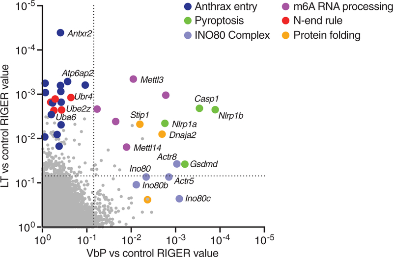
Intracellular pathogens and danger signals trigger the formation of inflammasomes, which activate inflammatory caspases and induce pyroptosis. The anthrax lethal factor metalloprotease and small-molecule DPP8/9 inhibitors both activate the NLRP1B inflammasome, but the molecular mechanism of NLRP1B activation is unknown. In this study, we used genome-wide CRISPR-Cas9 knockout screens to identify genes required for NLRP1B-mediated pyroptosis. We discovered that lethal factor induces cell death via the N-end rule proteasomal degradation pathway. Lethal factor directly cleaves NLRP1B, inducing the N-end rule-mediated degradation of the NLRP1B N terminus and freeing the NLRP1B C terminus to activate caspase-1. DPP8/9 inhibitors also induce proteasomal degradation of the NLRP1B N terminus but not via the N-end rule pathway. Thus, N-terminal degradation is the common activation mechanism of this innate immune sensor.


Figures

Fig. 1.
Fig. 1.. Genome-wide CRISPR-Cas9 screening identifies genes involved in NLRP1B-mediated pyroptosis.
Screens were performed in RAW 264.7 cells (see fig. S1). RIGER (RNAi gene enrichment ranking) values indicating the relative enrichment of genes after treatment with VbP (x axis) or LT (y axis) relative to control. The dotted lines indicate a RIGER p-value of 0.01.
Fig. 2.
Fig. 2.. LT and VbP induce proteasome-mediated degradation of NLRP1B.
(A) Diagram of NLRP1B. Single-letter abbreviations for the amino acid residues are as follows: F, Phe; K, Lys; L, Leu; M, Met; R, Arg; and S, Ser. (B and C) HEK 293T cells stably expressing mCasp1 (“m” denotes mouse) were transiently transfected with the indicated constructs (2 μg) for 24 hours, before cell viability was evaluated by lactate dehydrogenase (LDH) release (B) and expression was evaluated by immunoblotting (C). Residues that were mutated to create start sites are indicated. Data are means ± SEM of three biological replicates. ***P < 0.001 and **P < 0.01 by two-sided Student’s t test compared with mock. GST, glutathione S-transferase; GAPDH, glyceraldehyde phosphate dehydrogenase. (D) HEK 293T cells stably expressing mCasp1 were transiently transfected with a construct encoding GST-NLRP1B (30 ng). After 24 hours, cycloheximide (CHX, 100 mg/ml; used to block new protein synthesis), LT (1 μg/ml), and VbP (10 μM) were added to the indicated samples, which were then incubated for an additional 6 hours. FL, full-length. Asterisks indicate background bands. (E) HEK 293Tcells stably expressing mCasp1 were transiently transfected with a construct encoding V5-GFP-NLRP1B-FLAG (0.1 μg). After 24 hours, cells were treated with dimethyl sulfoxide (DMSO), bortezomib (Bort, 20 μM), MG-132 (20 μM), or Me-Bs (20 μM) for 30 min before the addition of either LT (1 μg/ml, 6 hours) or VbP (10 μM, 6 hours). Protein levels were evaluated by immunoblotting. (F) RAW 264.7 cells were treated with DMSO, bortezomib (20 μM), or MG-132 (20 μM) for 30 min before the addition of LT (1 μg/ml, 3 hours) or VbP (2 μM, 6 hours). Protein levels were evaluated by immune-blotting. Data are representative of three or more independent experiments.
Fig. 3.
Fig. 3.. The N-end rule pathway mediates LT-induced NLRP1B degradation.
(A) Knockout of Uba6, Ubr2, and Ubr4 in RAW 264.7 cells was confirmed by immunoblotting. (B to E) RAW 264.7 cells with the indicated genotypes were treated with LT (1 μg/ml) for 3 hours [(B) and (C)] or 6 hours [(D) and (E)] before supernatants were assessed for LDH release [(B) and (D)] and lysates were evaluated by immunoblotting [(C) and (E)]. (F) Knockout of both Ubr2 and Ubr4 in RAW 264.7 cells was confirmed by immunoblotting. (G and H) RAW 264.7 cells with the indicated genotypes were treated with LT (1 μg/ml) for 6 hours before supernatants were assessed for LDH release (G) and lysates were evaluated by immunoblotting (H). In (B), (D), and (G), data are means ± SEM of three biological replicates. *P < 0.05, **P < 0.01, ***P < 0.001, and NS (not significant) by two-sided Student’s t test. The same samples were used as controls in (D) and (G). (I) RAW 264.7 cells were treated with Me-Bs (10 μM) or L-Phe-NH2 (1 μM) for 30 min before the addition of LT (1 μg/ml, 6 hours). Supernatants were then evaluated for LDH release. Data are means ± SEM of three biological replicates. ***P < 0.001 by two-sided Student’s t test. (J and K) RAW 264.7 cells were treated with Me-Bs (10 μM), L-Phe-NH2 (1 μM), or bortezomib (1 μM) for 30 min before the addition of LT (1 μg/ml, 3 hours). Lysates were then evaluated by immunoblotting. Data are representative of three or more independent experiments.
Fig. 4.
Fig. 4.. The cleaved NLRP1B N terminus is sufficient to induce protein degradation.
(A) HEK 293T cells were transfected with the indicated small interfering RNAs, incubated for 24 hours, then transfected with NLRP1BM1−60- or NLRP1BL45−60-GFP-FLAG fusion constructs (0.05 μg) for an additional 24 hours. Lysates were then evaluated by immuno-blotting. (B) WT or UBR2 KO HEK 293T cells were transfected with the indicated NLRP1BM1−60- or NLRP1BL45−60-GFP-FLAG fusion constructs (0.5 μg) and UBR2 (1.5 μg) and incubated for 24 hours. Lysates were harvested, immunoprecipitated with anti-FLAG agarose beads, and evaluated by immunoblotting. Data are representative of three or more independent experiments. (C) Proposed model of LT-induced NLRP1B inflammasome activation.

Comment in

References

    1. Broz P, Dixit VM, Nat. Rev. Immunol. 16, 407–420 (2016). - PubMed
    1. Kayagaki N et al., Nature 526, 666–671 (2015). - PubMed
    1. Shi J et al., Nature 526, 660–665 (2015). - PubMed
    1. Levinsohn JL et al., PLOS Pathog. 8, e1002638 (2012). - PMC - PubMed
    1. Hellmich KA et al., PLOS ONE 7, e49741 (2012). - PMC - PubMed

Publication types

MeSH terms