. 2019 Apr 26;364(6438):eaav9011.
doi: 10.1126/science.aav9011. Epub 2019 Mar 14.

Architecture of the heteromeric GluA1/2 AMPA receptor in complex with the auxiliary subunit TARP γ8

Affiliations

Architecture of the heteromeric GluA1/2 AMPA receptor in complex with the auxiliary subunit TARP γ8

Beatriz Herguedas et al. Science. .

Abstract

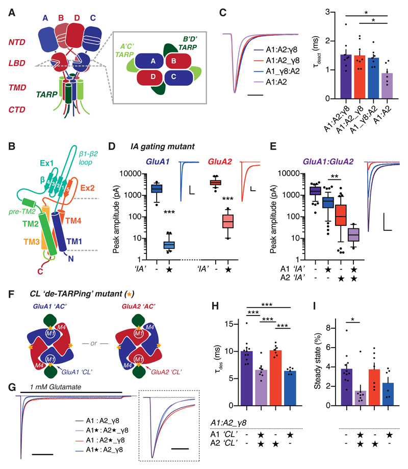
AMPA-type glutamate receptors (AMPARs) mediate excitatory neurotransmission and are central regulators of synaptic plasticity, a molecular mechanism underlying learning and memory. Although AMPARs act predominantly as heteromers, structural studies have focused on homomeric assemblies. Here, we present a cryo-electron microscopy structure of the heteromeric GluA1/2 receptor associated with two transmembrane AMPAR regulatory protein (TARP) γ8 auxiliary subunits, the principal AMPAR complex at hippocampal synapses. Within the receptor, the core subunits arrange to give the GluA2 subunit dominant control of gating. This structure reveals the geometry of the Q/R site that controls calcium flux, suggests association of TARP-stabilized lipids, and demonstrates that the extracellular loop of γ8 modulates gating by selectively interacting with the GluA2 ligand-binding domain. Collectively, this structure provides a blueprint for deciphering the signal transduction mechanisms of synaptic AMPARs.


Conflict of interest statement

Competing interests: The authors declare no competing interests.

Figures

Figure 1
Figure 1. Positioning of AMPAR subunits in the GluA1/2 heteromer.
(A) Schematic of an AMPAR tetramer (subunits A-D) depicting the four domain layers (NTD - N-terminal domain, LBD - ligand-binding domain, TMD - transmembrane domain, CTD - C-terminal domain). The AMPAR is two-fold symmetric with non-equivalent ‘pore-proximal’ (blue) and ‘pore-distal’ (red) positions. TARP auxiliary subunits can locate at two distinct sites denoted A’C’ (light green) or B’D’ (dark green). The inset (right) depicts the subunit arrangements as viewed from the LBD level. (B) Schematic of TARP γ8, depicting transmembrane helix (TM, cylinder) topology and arrangement of extracellular loops 1 and 2 (Ex1 & Ex2) into a beta sheet (arrows). (C) Deactivation kinetics of GluA1/2 receptors in response to 2 ms of 10 mM L-glutamate are slowed when γ8 is co-expressed or in tandem with the AMPAR, independently of the tandem fusion (deactivation τ (ms); A1:A2:γ8, 1.53 ± 0.18, n=8; A1:A2_γ8, 1.51 ± 0.16, n=8; A1_γ8:A2, 1.43 ± 0.12, n=7; A1:A2, 0.89 ± 0.14, n=6; One-way ANOVA with Tukey’s multiple comparison test, p=0.0316). Peak-scaled responses, scale bar = 3 ms. (D) GluA1 I629A and GluA2 I633A mutations (denoted ‘IA’, or starred) impair current responses of homomeric receptors (10 mM L-glutamate peak amplitude (pA) (median, 25 and 75 percentiles reported, 5 to 95 percentiles depicted as whiskers); GluA1, 1991.3 (1323.5 to 3214.8), n=19; GluA1 I629A, 5.1 (3.3 to 7.1), n=22; Mann-Whitney test, p<0.0001; GluA2, 3932.0 (2989.0, 5080.9), n=20; GluA2 I633A, 59.9 (29.7, 129.1), n=19; Mann-Whitney test, p<0.0001). Scale bars = 400 pA & 20 ms. (E) In a GluA1/2 heteromeric context, GluA2 IA mutation impairs channel gating significantly more than GluA1 IA (Peak amplitude (pA); GluA1/2, 1520.9 (1017.5, 2467.3), n=48; GluA1IA/A2, 538.2 (254.2, 823.3), n=48; GluA1/A2IA, 103.5 (38.4, 366.6), n=42; GluA1IA/A2IA, 14.5 (7.2, 29.7), n=18; Kruskal-Wallis test with Dunn’s multiple comparisons test, p<0.0001, all conditions are significantly different to each other, however only statistical significance of A1IA/A2 versus A1/A2IA comparison is shown for clarity), demonstrating preferential pore-distal location of GluA2 in the heteromeric receptor. Scale bar = 20 ms & 400 pA. (F) ‘CL’ mutation of M1 helix (yellow star) abolishes TARP-modulation of AMPAR gating. Two subunit arrangements ‘GluA1 pore-proximal’ and ‘GluA2 pore-proximal’ are possible. In a GluA1/2_γ8 receptor, mutagenesis will prevent TARP-modulation when included in pore-proximal, but not pore-distal subunits. (G) Sample traces for 500 ms application of 1 mM L-glutamate to A1/A2_γ8 with or without ‘CL’ mutation (star). Peak-scaled responses, scale bar = 100 ms, and magnified inset scale bar = 10 ms. (H) TARP-dependent slowing of desensitization is prevented by CL mutation of GluA1 (C524L), but not GluA2 (C528L) (desensitization τ (ms); A1:A2_γ8, 10.1 ± 0.6, n=11; A1CL:A2CL_γ8, 6.6 ± 0.5, n=8; A1:A2CL_γ8, 10.2 ± 0.4, n=7; A1CL:A2_γ8, 6.4 ± 0.2, n=6; one-way ANOVA with Tukey’s multiple comparison test, p<0.0001), which is reflected in the magnitude of steady-state AMPAR currents (I) (Steady-state response as % of peak amplitude; A1:A2_γ8, 3.82 ± 0.49, n=11; A1CL:A2CL_γ8, 1.55 ± 0.59, n=8; A1:A2CL_γ8, 3.76 ± 0.67, n=7; A1CL:A2_γ8, 2.37 ± 0.57, n=6; One-way ANOVA with Tukey’s multiple comparison test, p=0.02).
Figure 2
Figure 2. Cryo-EM structure of the full-length GluA1/2_γ8 receptor complex.
(A) Cryo-EM density map of the full-length GluA1/2_γ8 receptor (B-factor sharpening = -350). The structural model was obtained by rigid-body fitting of individual receptor domains into the density map (GluA1 in blue, GluA2 in red, γ8 in green). A top view onto the ‘NTD layer’ is shown above the receptor, highlighting the tetramer interface formed by the two GluA2 NTDs. (B) Model depicting front view of the full-length GluA1/2_γ8 heteromer. The three domain layers, NTD, LBD and TMD, are indicated, and stippled lines indicate the boundaries of the membrane. (C) Side view of the receptor model, visualizing the tetrameric interface in the NTD layer, a GluA1/GluA2 LBD heterodimer and two γ8 auxiliary subunits docked at the B’D’ positions. (D) Slice of the 3D cryo-EM map at the TMD region showing transmembrane helices of GluA1 (blue), GluA2 (red) and γ8 (green). (E) Detail of the cryo-EM density at the GluA1-specific glycosylation site on the NTD. At A/C subunits, protruding density corresponds to a GluA1-specific glycan site (Asn45). (F) The B/D subunit (GluA2) shows no extra density for a glycan.
Figure 3
Figure 3. Cryo-EM map of the GluA1/2_γ8 TMD-LBD region.
(A) Cryo-EM map (4.4 Å) of the TMD/LBD/γ8 portion of the A1/2_γ8 complex (B-factor sharpening = -150) with GluA1 (blue), GluA2 (red) and γ8 (green) showing lipid-like densities demarcating the upper membrane boundary (orange). Color-scheme is maintained throughout the figure. The competitive antagonist NBQX is located in the agonist-binding site of the LBD. (B) Top view of the TMD-LBD map showing the two LBD heterodimers forming the ‘gating ring’ in a characteristic two-fold symmetric “closed-state” conformation. TARP γ8 is positioned beneath GluA2 subunits. (C) Detail of the M3 gate helices of the TMD region, below the LBD layer, highlighting the position of GluA1 I629 and GluA2 I633 (spheres). (D) Details of the selectivity filter forming the inner pore constriction. Side view onto the filter showing the Q/R site residues on top of the pore loop and the adjacent +1 Q (GluA1 Q583, GluA2 Q587). The cysteines at the +3 position (GluA1 C585, GluA2 C589) surround a strong density within the pore. The polyamine-binding aspartates (GluA1 D586, GluA2 D590) are also indicated. The selectivity filter is surrounded by the M3 gating helices. (E) Top view, showing the orientation of the Q/R site residues (GluA1 Q582, GluA2 R586) and the +1Q side chains interacting with tryptophans projecting from the M3 helices (GluA1 W602, GluA2 W606).
Figure 4
Figure 4. Lipid-like densities associate with the TMD region of GluA1/2_γ8.
(A) Model of the TMD region (as cartoon) with five modeled acyl chains (L1 to L5, as sticks in orange) filling the cavity between γ8 TM4 and GluA1 M4, and a further lipid molecule (L6) locating to the perimeter of γ8, between the γ8 TM1 and TM2 helices. Cryo-EM density for lipid-like molecules is shown as a mesh. (B) Detailed view of lipids L4 and L5. The density for L4 projects into the fenestration between M1 and M3, approaching the apex of the M2 loop of the GluA1 subunit. Side chains interacting with the potential lipid molecules are represented as sticks. (C) TMD slice (right) of the cryo-EM map of the TMD/LBD model (left), with clear density for the GluA1, GluA2 and γ8 helices and the 6 acyl groups color-coded as in (A). (D) Bottom view of the A1/2_γ8 TMD sector with the modeled lipids displayed as orange spheres.
Figure 5
Figure 5. Interaction of γ8 with the GluA1/2 TMD region.
(A) Ribbon representation of the A1/2_γ8 TMD-LBD model. Numbers and dashed-lines represent the points of view of the AMPAR-γ8 interaction site as displayed in panels (B), (C) and (D). (B) Top view of the AMPAR-γ8 interaction region, with the cryo-EM map overlayed. Important interacting residues are shown as sticks. This slice through the density was generated at the level of γ8 residues V176 (TM3) and G209 (TM4) to offer a view onto the ‘pocket’ occupied by antiepileptic drugs. (C) Side view of the interaction between γ8 (green) and the receptor. Docking of the TARP results in helical reorganization, highlighted by overlaying M1 of the TARP-free (A’C’) site (grey helices) with the TARP-bound B’D’ site (colored helices). The helical shift between M4 helices (~1.5 Å) is shown. A bottom view is depicted inset. (D) TARP binding results in M1 side chain reorganizations (obtained by overlaying M1 helices from the A’C’ and B’D’ sites, as described in panel (C). GluA1 Y519 and M523 move to interact with the TARP, while E520 forms a hydrogen bond with Y199 of γ8 (depicted in magnified inset). (E) Side view of the interaction between γ8 TM3 helix (green) and GluA2 M4 helix (red), with interacting side chains displayed as sticks and contoured with the cryo-EM density. (F) Interaction between γ8 TM4 helix (green) and GluA1 M1 helix (blue), with interacting side chains displayed as sticks and contoured with the cryo-EM density.
Figure 6
Figure 6. A unique TARP γ8 interaction site on the GluA2 LBD controls receptor gating.
(A) Full-length 2 cryo-EM map showing the additional density between the upper lobe of GluA2 and the γ8 β1-β2 loop in Ex1 (represented with B= -170 and using a low-threshold). Magnified inset contains TMD/LBD model as a cartoon representation, showing GluA2 (red) and γ8 (green). (B) Surface representation of the TMD/LBD model, depicting the likely location of β1-β2 loop residues (dashed line) projecting towards the GluA2 LBD upper lobe loop. Location of the glyco-mutation for disruption of this interaction is depicted as a star. (C) Current responses of TARP-associated A1/2 receptors evoked by 500 ms 1 mM L-glutamate application both with (light green) and without (dark green) GluA2 E413T (glyco-) mutation to introduce N-glycosylation (on N411) at the predicted interaction site. Glycosylation selectively affects the rate of entry into desensitization of TARP γ8 but not TARP γ2-associated A1/2 receptors. Peak-scaled responses, scale bar = 10 ms. (D) GluA2 glyco-mutation speeds the rate of desensitization entry of A1/2_γ8 to that of TARP-free A1/2 receptors. In contrast, glyco-mutation of GluA1 in A1/2_γ8 or GluA2 in an A1/2_γ2 assembly do not affect desensitization entry kinetics (desensitization τ (ms); A1/A2 (grey bar), 6.27 ± 0.33, n=5; A1/A2_γ8, 9.68 ± 0.46, n=25; A1/A2glyco_γ8, 7.36 ± 0.30, n=15; A1glyco/2_γ8, 10.71 ± 0.51, n=5; A1/A2_γ2, 10.14 ± 0.72, n=10; A1/A2glyco_γ2, 9.71 ± 1.05, n=6; One-way ANOVA with Sidak’s multiple comparison test, p=0.0005).

References

    1. Greger IH, Watson JF, Cull-Candy SG. Structural and Functional Architecture of AMPA-Type Glutamate Receptors and Their Auxiliary Proteins. Neuron. 2017;94:713–730. - PubMed
    1. Traynelis SF, et al. Glutamate receptor ion channels: structure, regulation, and function. Pharmacol Rev. 2010;62:405–496. - PMC - PubMed
    1. Huganir RL, Nicoll RA. AMPARs and synaptic plasticity: the last 25 years. Neuron. 2013;80:704–717. - PMC - PubMed
    1. Sommer B, Kohler M, Sprengel R, Seeburg PH. RNA editing in brain controls a determinant of ion flow in glutamate-gated channels. Cell. 1991;67:11–19. - PubMed
    1. Hume RI, Dingledine R, Heinemann SF. Identification of a site in glutamate receptor subunits that controls calcium permeability. Science. 1991;253:1028–1031. - PubMed

Publication types

Substances

LinkOut - more resources