. 2019 Mar 14;9(1):4479.
doi: 10.1038/s41598-019-39617-0.

Corneal remodelling and topography following biological inlay implantation with combined crosslinking in a rabbit model

Affiliations

Corneal remodelling and topography following biological inlay implantation with combined crosslinking in a rabbit model

Iben Bach Damgaard et al. Sci Rep. .

Abstract

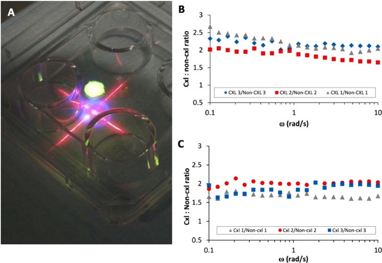
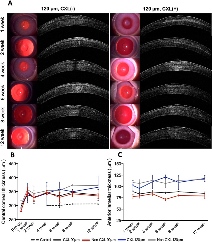
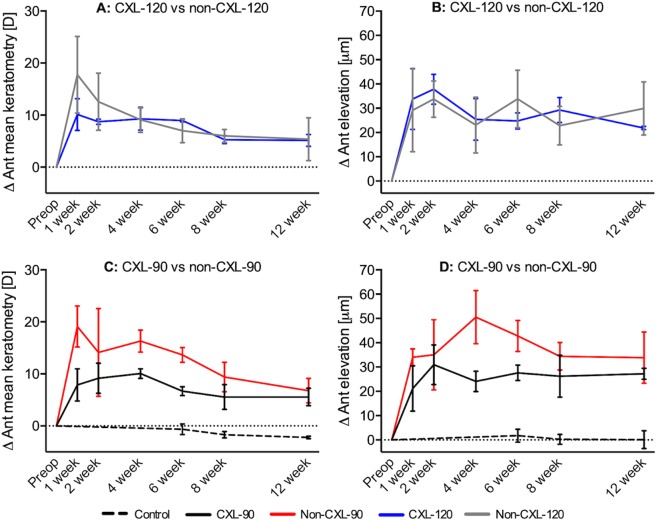
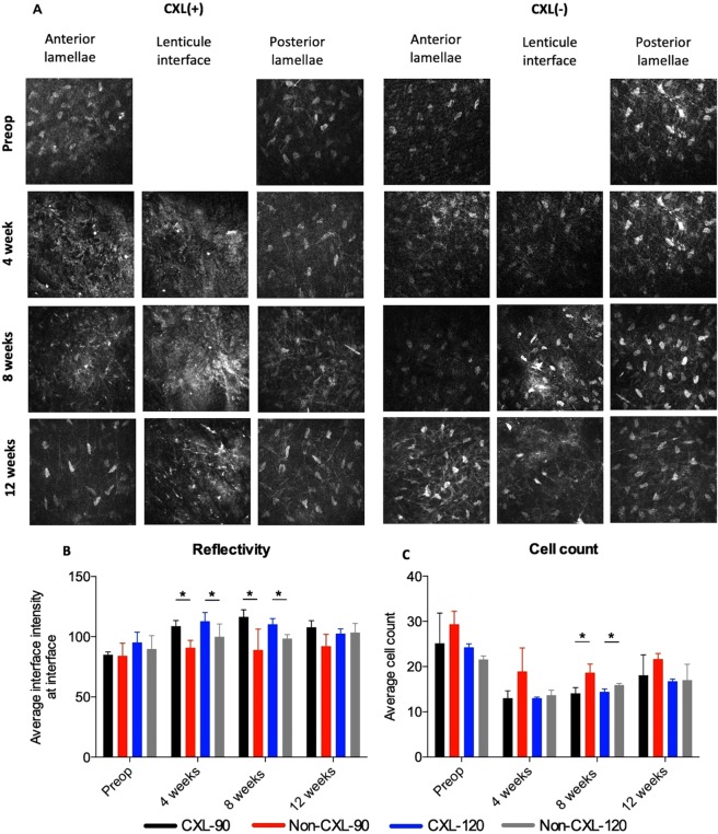
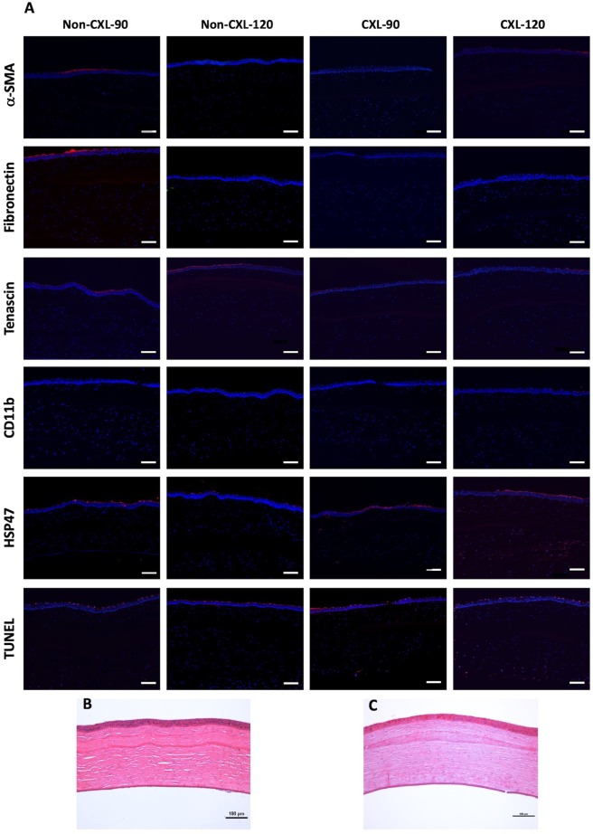
Implantation of biological corneal inlays, derived from small incision lenticule extraction, may be a feasible method for surgical management of refractive and corneal diseases. However, the refractive outcome is dependent on stromal remodelling of both the inlay and recipient stroma. This study aimed to investigate the refractive changes and tissue responses following implantation of 2.5-mm biological inlays with or without corneal collagen crosslinking (CXL) in a rabbit model. Prior to implantation, rotational rheometry demonstrated an almost two-fold increase in corneal stiffness after CXL. After implantation, haze gradually subsided in the CXL-treated inlays (p = 0.001), whereas the untreated inlays preserved their clarity (p = 0.75). In-vivo confocal microscopy revealed reduced keratocyte cell count at the interface of the CXL inlays at week 8. Following initial steepening, regression was observed in anterior mean curvature from week 1 to 12, being most prominent for the non-CXL subgroups (non-CXL: -12.3 ± 2.6D vs CXL: -2.3 ± 4.4D at 90 μm depth, p = 0.03; non-CXL: -12.4 ± 8.0D vs CXL: -5.0 ± 4.0D at 120 μm depth, p = 0.22). Immunohistochemical analysis revealed comparable tissue responses in CXL and untreated subgroups. Our findings suggest that CXL of biological inlays may reduce the time before refractive stabilization, but longer postoperative steroid treatment is necessary in order to reduce postoperative haze.


Conflict of interest statement

Dr. Mehta is a consultant for Ziemer and Carl Zeiss Meditec, Inc. The remaining authors have no conflicts of interests.

Figures

Figure 1
Figure 1
Comparison of biomechanical strength between non-CXL and CXL lenticules assessed by rheometry. (A) Corneal collagen crosslinking of a rabbit lenticule. A central 2.5-mm button was trephined after CXL and used for implantation. Frequency dependent crosslinked lenticule/non-crosslinked lenticule G’ ratio for (B) human lenticules and (C) rabbit lenticules.
Figure 2
Figure 2
Slit lamp observation and anterior segment-optical coherence tomography (AS-OCT) of corneas following implantation of CXL and untreated inlays. (A) Slit-lamp biomicroscopy and anterior segment optical coherence tomography (ASOCT). Left column: Untreated inlays. The inlay preserved their stromal clarity during the postoperative period. Right column: CXL treated inlays. The postoperative stromal haze progressively improved from week 1 to week 12. (B) The development in the average central corneal thickness and (C) the anterior lamellar thickness. Bars represents standard deviations.
Figure 3
Figure 3
Refractive changes following implantation of non-CXL and CXL inlays at different corneal depths. The average change in (A,C) anterior mean curvature and (B,D) anterior elevation at different time points during postoperative follow up. Bars represents standard deviations.
Figure 4
Figure 4
In vivo confocal microscopy of corneal stroma following implantation of non-CXL and CXL inlays. (A) Representative in vivo confocal micrographs of the anterior stroma, inlay interface and posterior stroma. (B) The average intensity measured in mean gray value (MGV) using a semi-automated tracking program (ImageJ). (C) Average keratocyte cell count evaluated by the mean of four representative images of the interface. Bars represent standard deviations. *Significant difference, p < 0.05.
Figure 5
Figure 5
Tissue responses following implantation of non-CXL and CXL inlays in rabbit corneas. (A) Immunohistochemistry assays of α-SMA, fibronectin, tenascin, CD11b, heat shock protein 47 (HSP47), and TUNEL 12 weeks postoperatively. Nuclei are counterstained using DAPI (blue). All images were captured at x100 magnification; scale bars represent 100 µm. (B + C) Representative hematoxylin-eosin staining images for CXL treated inlays at (B) 90 μm and (C) 120 μm depth. Original magnification: 100x. Scale bars represent 100 µm.

References

    1. Strenk SA, et al. Age-related changes in human ciliary muscle and lens: a magnetic resonance imaging study. Invest. Ophthalmol. Vis. Sci. 1999;40:1162–9. - PubMed
    1. Konstantopoulos A, Mehta JS. Surgical compensation of presbyopia with corneal inlays. Expert Rev. Med. Devices. 2015;12:341–52. doi: 10.1586/17434440.2015.1007124. - DOI - PubMed
    1. Dexl AK, et al. Long-term outcomes after monocular corneal inlay implantation for the surgical compensation of presbyopia. J. Cataract Refract. Surg. 2015;41:566–575. doi: 10.1016/j.jcrs.2014.05.051. - DOI - PubMed
    1. Jalali S, Aus der Au W, Shaarawy T. AcuFocus Corneal Inlay to Correct Presbyopia Using Femto-LASIK. One Year Results of a Prospective Cohort Study. Klin. Monbl. Augenheilkd. 2016;233:360–364. doi: 10.1055/s-0041-111804. - DOI - PubMed
    1. Ismail MM. Correction of hyperopia by intracorneal lenses. Two-year follow-up. J. Cataract Refract. Surg. 2006;32:1657–1660. doi: 10.1016/j.jcrs.2005.08.057. - DOI - PubMed

Publication types