. 2019 Mar 14;9(1):4582.
doi: 10.1038/s41598-019-40820-2.

An analysis of benign human prostate offers insights into the mechanism of apocrine secretion and the origin of prostasomes

Affiliations

An analysis of benign human prostate offers insights into the mechanism of apocrine secretion and the origin of prostasomes

Nigel J Fullwood et al. Sci Rep. .

Abstract

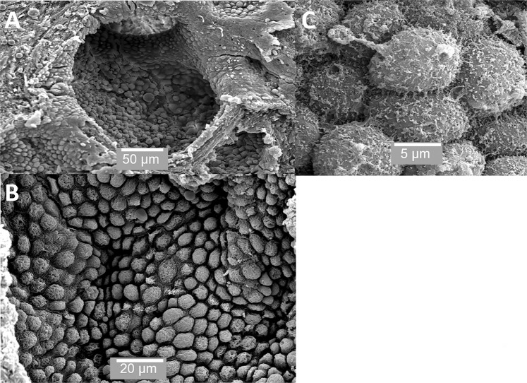
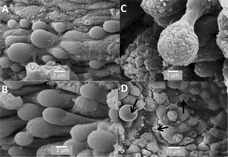
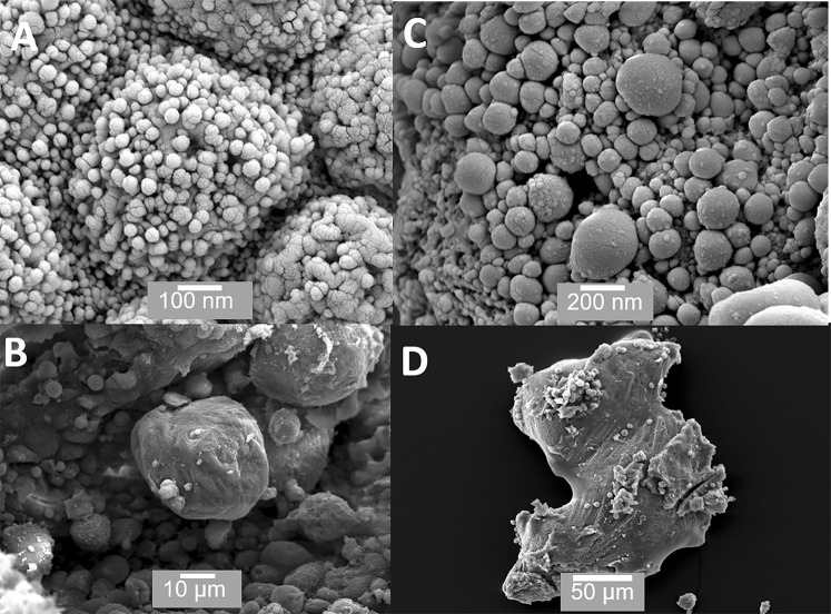
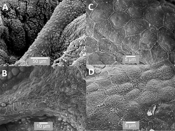
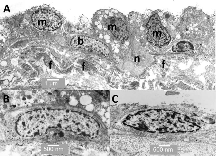
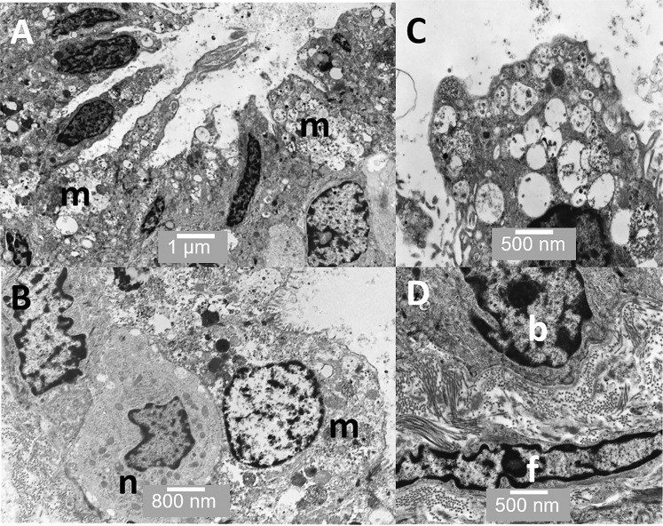
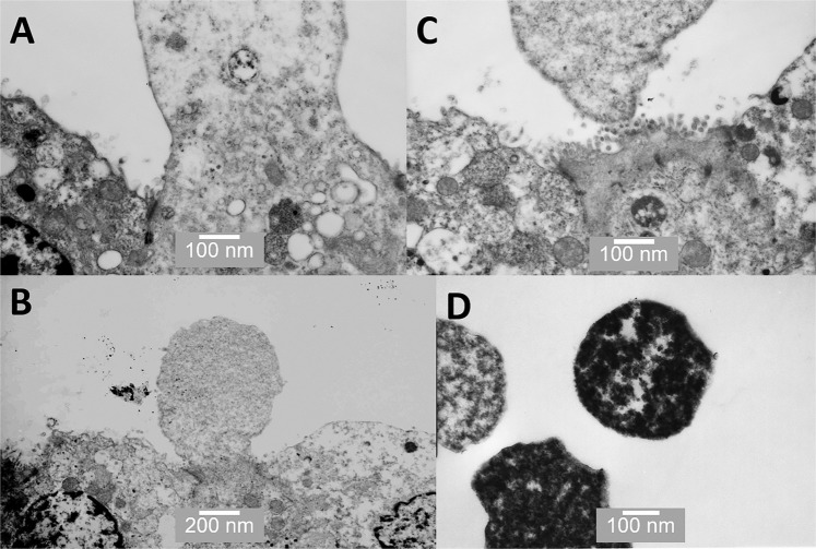
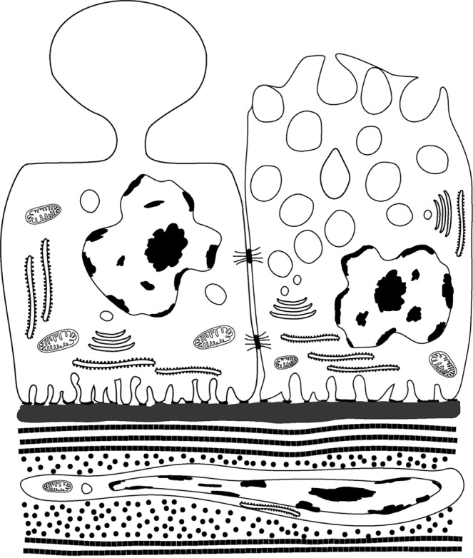
The structure and function of normal human prostate is still not fully understood. Herein, we concentrate on the different cell types present in normal prostate, describing some previously unreported types and provide evidence that prostasomes are primarily produced by apocrine secretion. Patients (n = 10) undergoing TURP were prospectively consented based on their having a low risk of harbouring CaP. Scanning electron microscopy and transmission electron microscopy was used to characterise cell types and modes of secretion. Zinc levels were determined using Inductively Coupled Plasma Mass Spectrometry. Although merocrine secretory cells were noted, the majority of secretory cells appear to be apocrine; for the first time, we clearly show high-resolution images of the stages of aposome secretion in human prostate. We also report a previously undescribed type of epithelial cell and the first ultrastructural image of wrapping cells in human prostate stroma. The zinc levels in the tissues examined were uniformly high and X-ray microanalysis detected zinc in merocrine cells but not in prostasomes. We conclude that a significant proportion of prostasomes, possibly the majority, are generated via apocrine secretion. This finding provides an explanation as to why so many large proteins, without a signal peptide sequence, are present in the prostatic fluid.


Conflict of interest statement

The authors declare no competing interests.

Figures

Figure 1
Figure 1
SEM of prostate. (A) shows an individual acinus; (B) shows the considerable variation in the size of the cells from 10 μm to 5 μm and (C) shows the surface of individual cells. Scale bars: A - 50 µm, B - 20 µm, C - 5 µm.
Figure 2
Figure 2
SEM of prostate showing cells undergoing apocrine secretion. (A,B) shows the apical surface of the cells to consist of rounded dome-like structures; (C) shows the budding off process; (D) shows completely detached membrane-bound aposomes (arrows), some over a micron in size. Scale bars: A - 2 µm, B - 2 µm, C - 1 µm, D - 10 µm.
Figure 3
Figure 3
SEM of material found within acini. (A) Small (<50 nm) vesicles or exosomes are sometimes visible on the surface of cells; (B) shows a small rounded prostatic calculi; (C) shows the interior of an acinus packed with prostasomes ranging in size from 150 nm to several hundred nanometres; (D) shows an irregular prostatic calculi >200 μm in size (D). Scale bars: A - 100 nm, B - 10 µm, C - 200 nm, D - 50 µm.
Figure 4
Figure 4
SEM of the ridge-like structures, which separate adjacent acini. (A,B) The cells running along the ridge-like structures appear different to those within the acini; and, (C,D) shows the cells to be flat and polygonal with very clearly defined borders. Scale bars: A - 50 µm, B - 10 µm, C - 5 µm, D - 5 µm.
Figure 5
Figure 5
TEM through an acinus. (A) Several merocrine secretory cells are evident (m) as well as a basal cell (b) and a neuroendocrine cell (n). Collagen fibrils are also visible (f) (B) shows a basal cell in more detail; and (C) shows a cuboid epithelial-like cell from the ridge region between the acini. Scale bars: A - 1 µm, B - 500 nm, C - 500 nm.
Figure 6
Figure 6
TEM through an acinus. (A) Shows several highly active merocrine secretory cells (m); (B) shows a neuroendocrine cell (n) below a merocrine secretory cell (m); (C) shows the apical region of one of the merocrine secretory cells filled with vesicles; and (D) shows a basal cell (b) and a fibroblast (f) in the stroma immediately below it. Scale bars: A - 1 µm, B - 800 nm, C - 500 nm, D - 500 nm.
Figure 7
Figure 7
TEM images of various stages of apocrine secretion. (A) shows the protrusion of a dome from the apical surface; (B) shows the narrowing at the base of the dome to form a neck; (C) shows the separation of the aposome from the cell surface; and (D) shows the separated aposomes clearly containing some dense cytoplasmic material. Scale bars: A - 100 nm, B - 200 nm, C - 100 nm, D - 100 nm.
Figure 8
Figure 8
TEM images of the prostate stroma. (A) shows a minor blood vessel lined with vascular endothelial cells (v); (B) shows smooth muscle in longitudinal section, note the dense bodies (arrowheads) found in smooth muscle; (C) shows smooth muscle in cross-section surrounded by collagen bundles; and (D) shows a wrapping cell (w) surrounding a muscle bundle (s). Scale bars: A - 2 µm, B - 500 nm, C - 500 nm, D - 500 nm.
Figure 9
Figure 9
The illustration shows the differences between apocrine (left) and merocrine (right) secretory cells. Apocrine cells are characterised by dome-like protrusions, which pinch off and are released into the lumen. The vesicles in merocrine secretory vesicles are derived from the RER and Golgi apparatus and the contents released via the fusing with the apical membrane and exocytosis. The bottom of the illustration shows a sub-epithelial stromal fibroblast surrounded by collagen fibrils.
Figure 10
Figure 10
This micrograph orientation figure shows a greatly simplified diagram of a 2-D cross section through the centre of an acini with 3-D symmetry about the yellow line. The perspective of the TEM 2-D micrographs is the same as in this 2-D diagram. The perspective of the SEM micrographs should be visualised as being from the origin of the dotted arrows. Figures 1, 2, 3, 5A,B, 6 and 7 are from the area labelled 1. Figures 4 and 5C are from the areas labelled 2. Figure 8 is from the area labelled 3. Secretory cells are shown as red, putative basal stem cells as blue and the non-secretory cuboid epithelial cells as purple.

References

    1. Zhou CK, et al. Prostate cancer incidence in 43 populations worldwide: An analysis of time trends overall and by age group. Int. J. Cancer. 2016;138:1388–1400. doi: 10.1002/ijc.29894. - DOI - PMC - PubMed
    1. Martin FL, et al. Constitutive expression of bioactivating enzymes in normal human prostate suggests a capability to activate pro-carcinogens to DNA-damaging metabolites. Prostate. 2010;70:1586–1599. doi: 10.1002/pros.21194. - DOI - PubMed
    1. Theophilou G, et al. A biospectroscopic analysis of human prostate tissue obtained from different time periods points to a trans-generational alteration in spectral phenotype. Sci. Rep. 2015;5:13465. doi: 10.1038/srep13465. - DOI - PMC - PubMed
    1. McNeal JE. Normal histology of the prostate. Am. J. Surg. Pathol. 1988;12:619–633. doi: 10.1097/00000478-198808000-00003. - DOI - PubMed
    1. Ragavan N, et al. CYP1B1 expression in prostate is higher in the peripheral than in the transition zone. Cancer Lett. 2004;215:69–78. doi: 10.1016/j.canlet.2004.06.051. - DOI - PubMed

Publication types