. 2019 Mar 14;9(1):4603.
doi: 10.1038/s41598-019-40922-x.

Extended insight into the Mycobacterium chelonae-abscessus complex through whole genome sequencing of Mycobacterium salmoniphilum outbreak and Mycobacterium salmoniphilum-like strains

Affiliations

Extended insight into the Mycobacterium chelonae-abscessus complex through whole genome sequencing of Mycobacterium salmoniphilum outbreak and Mycobacterium salmoniphilum-like strains

Phani Rama Krishna Behra et al. Sci Rep. .

Abstract

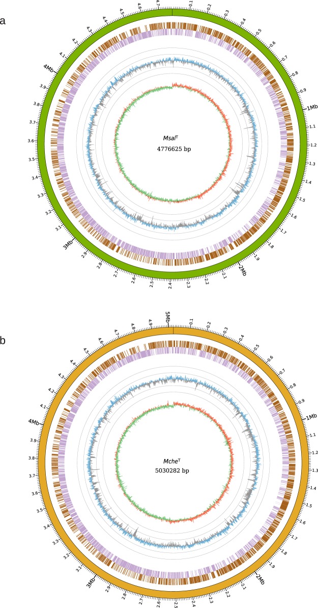
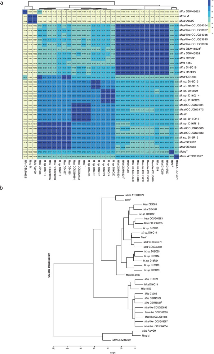
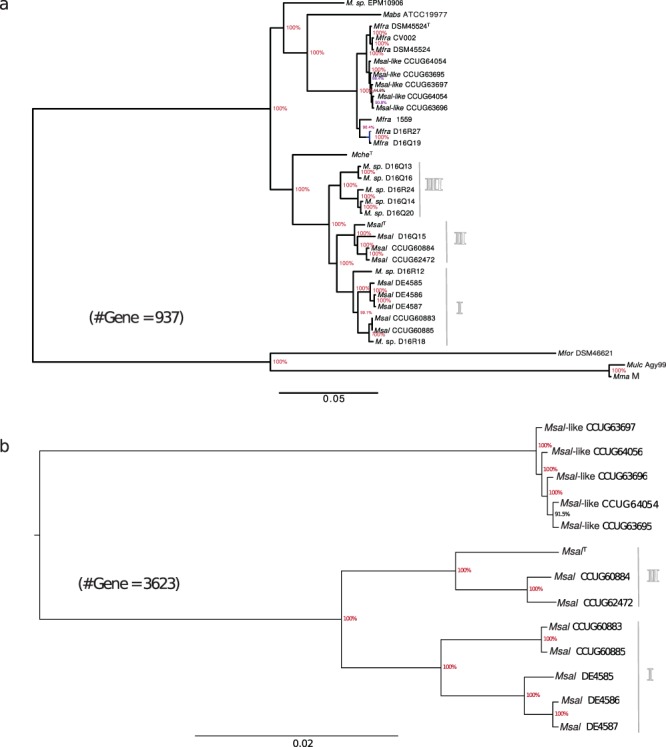
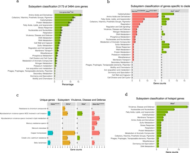
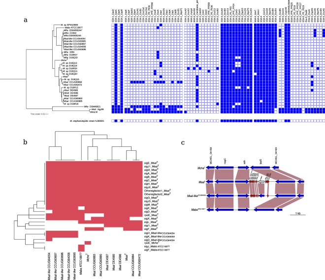
Members of the Mycobacterium chelonae-abscessus complex (MCAC) are close to the mycobacterial ancestor and includes both human, animal and fish pathogens. We present the genomes of 14 members of this complex: the complete genomes of Mycobacterium salmoniphilum and Mycobacterium chelonae type strains, seven M. salmoniphilum isolates, and five M. salmoniphilum-like strains including strains isolated during an outbreak in an animal facility at Uppsala University. Average nucleotide identity (ANI) analysis and core gene phylogeny revealed that the M. salmoniphilum-like strains are variants of the human pathogen Mycobacterium franklinii and phylogenetically close to Mycobacterium abscessus. Our data further suggested that M. salmoniphilum separates into three branches named group I, II and III with the M. salmoniphilum type strain belonging to group II. Among predicted virulence factors, the presence of phospholipase C (plcC), which is a major virulence factor that makes M. abscessus highly cytotoxic to mouse macrophages, and that M. franklinii originally was isolated from infected humans make it plausible that the outbreak in the animal facility was caused by a M. salmoniphilum-like strain. Interestingly, M. salmoniphilum-like was isolated from tap water suggesting that it can be present in the environment. Moreover, we predicted the presence of mutational hotspots in the M. salmoniphilum isolates and 26% of these hotspots overlap with genes categorized as having roles in virulence, disease and defense. We also provide data about key genes involved in transcription and translation such as sigma factor, ribosomal protein and tRNA genes.


Conflict of interest statement

The authors declare no competing interests.

Figures

Figure 1
Figure 1
Overview of the MsalT and McheT genomes. (a) Circos plot showing the complete genome sequence of MsalT. From outer to inner circle: Green track represents the complete genome overlapping with scale along the genome length. The next two circles, marked as brown and violet blocks, represent genes in forward (brown) and reverse (violet) strands. The circle with blue (higher than the mean value) and grey (lower than the mean value) “spikes” show the GC-content distribution calculated using a sliding window of 1000 bp, while each grey circle represent variations of the mean GC-content 64.3% in ±10 and ±20 units (i.e. outer grey circle = 84.3% and inner grey circle = 44.3%). The inner track in red (positive) and green (negative) circle shows the GC-skew using a sliding window of 1000 bp. (b) Same as in (a) for McheT where the complete genome overlapping with scale along the genome length (outer circle), which is illustrated in dark yellow. The mean GC-value equals to 63.9%.
Figure 2
Figure 2
Clustering of Msal, Msal-like and other MCAC-members based on the average nucleotide scores (ANI) as indicated. (a) Heat map showing ANI values for ‘all-versus-all’ Msal and Msal-like strains including other members of the MCAC. ANI values were clustered based on unsupervised hierarchical clustering (see Methods). The horizontal tree represents the heatmap clustering of column wise dendogram. (b) Dendogram, extracted from the heat map in (a), showing clustering of different strains/isolates based on ANI values.
Figure 3
Figure 3
Comparative analysis of orthologous genes predicted to be present in Msal and Msal-like strains, McheT and MabsATCC19977. (a) Circos plot showing the presence of protein coding genes in seven Msal, five Msal-like strains, McheT and MabsATCC19977 compared to the reference genome MsalT. The outer track (green) represents the genome for MsalT with a size scale, while the next circle in blue corresponds to the predicted protein coding sequences (CDS) for MsalT. Subsequent circular tracks represent one genome and the number corresponds to the strain name in the legend on the right. Colored radial blocks represent orthologous genes in the corresponding genome and color intensity (see color scale in the middle) indicates percentage identity at the protein level. The white blocks indicate that no orthologs were identified. (b) Heat map showing presence (red) and absence (white) of orthologous genes (excluding core genes) mapped in different Msal and Msal-like strains, McheT and MabsATCC19977, and clustered using hierarchical clustering. The horizontal and vertical trees represent the heat map clustering of the column and row wise dendograms. (c) Venn diagram showing common and unique coding genes for Msal and Msal-like representative strains as indicated. (d) Venn diagram showing common and unique genes in MsalT, Msal-likeCCUG64054, McheT and MabsATCC19977. (e) Heat map showing presence (red) and absence (white) of orthologous ncRNA genes mapped as in (b). The ncRNA genes were identified using Rfam, see main text. The horizontal and vertical trees represent the heat map clustering of the column and row wise dendograms.
Figure 4
Figure 4
Phylogenetic relationship of Msal and Msal-like strains, McheT and MabsATCC19977. (a) Phylogenetic tree based on 937 core genes present in Msal, Mfra (Msal-like) strains, McheT, MabsATCC19977, MforDSM46621, MulcAgy99 and Mma (M strain). For details see Methods. (b) Phylogenetic tree based on 3623 core genes present in Msal and Msal-like strains as indicated.
Figure 5
Figure 5
Functional classification of genes in MsalT, Msal-likeCCUG64054, McheT and MabsATCC19977 into subsystem as indicated. (a) Subsystem classification of 2173 core genes using MsalT. Of note, that a gene can be classified in more than one subsystem. (b) Subsystem classification of specific genes present in MfraCCUG64054 and MabsATCC19977, and present in MsalT and McheT as indictated. (c) Classification of unique genes present in MabsATCC19977, McheT, MsalT and Msal-likeCCUG64054 in the subsystem “Virulence, Disease and Defence”. (d) Subsystem classification of mutational hotspot genes in Msal, see main text for details.
Figure 6
Figure 6
Analysis of ESX, sigma factor and tRNA genes in MCAC-members. (a) ESX related genes. Presence (blue) and absence (white) of ESX related genes in different mycobacteria as indicated in the phylogenetic tree shown to left (see also Fig. 4b). ESX related genes present in M. stephanolepidis NJB0901 is shown below, see main text. (b) Sigma factor genes. Heat map showing presence (red) and absence (white) of sigma factor genes in MabsATCC19977, McheT, MsalT and Msal-likeCCUG64054. The signature for the respective sigma factor genes correlate with the naming for MtbH37Rv sigma factor genes. The horizontal and vertical trees represent the heat map clustering of the column and row wise dendograms. (c) Predicted presence of additional tRNA genes in MCAC-members. Gene synteny for a tRNA gene cluster encompassing nine genes in MsalT (seven in MfraCCUG64054). The tRNA genes are marked in red and the vertical boxes marked in brown highlight homologous genes. Note the presence of the HNH endonuclease gene (marked in gray) located within tRNA gene clusters (see main text). See also Figs S1a and S9a.

References

    1. Bataillon E, Dubard TL. Un nouveau type de tuberculose. Comptes rendus des Sceances de la Societe Biologie. 1897;49:446–449.
    1. Austin, B. & Austin, D. A. Bacterial Fish Pathogens (SpringerDordrecht Heidelberg New York London) (2013).
    1. Whitman, W. B. et al. Bergey’s Manual® of Systematic Bacteriology. 2nd ed. Springer New York: New York, NY (2012).
    1. Righetti M, et al. Mycobacterium salmoniphilum infection in a farmed Russian sturgeon, Acipenser gueldenstaedtii (Brandt & Ratzeburg) J Fish Dis. 2014;37:671–674. - PubMed
    1. Zerihun MA, Nilsen H, Hodneland S, Colquhoun DJ. Mycobacterium salmoniphilum infection in farmed Atlantic salmon, Salmo salar L. J Fish Dis. 2011;34:769–781. - PubMed

Publication types

Substances