. 2019 Mar 14;9(1):4452.
doi: 10.1038/s41598-019-41051-1.

Steroidogenic Factor 1 (Nr5a1) is Required for Sertoli Cell Survival Post Sex Determination

Affiliations

Steroidogenic Factor 1 (Nr5a1) is Required for Sertoli Cell Survival Post Sex Determination

Prashanth Anamthathmakula et al. Sci Rep. .

Abstract

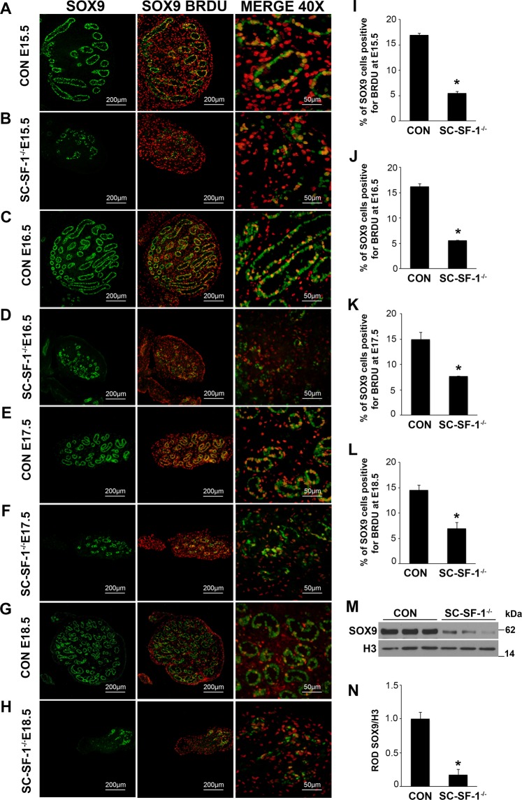
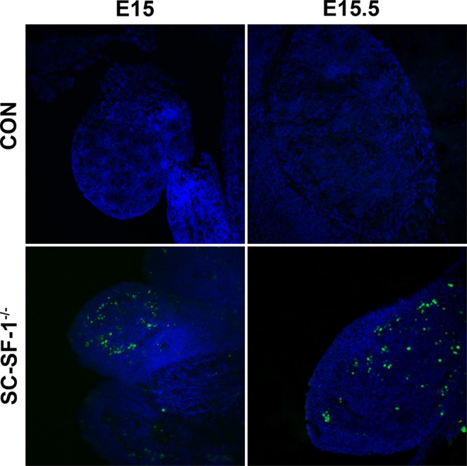
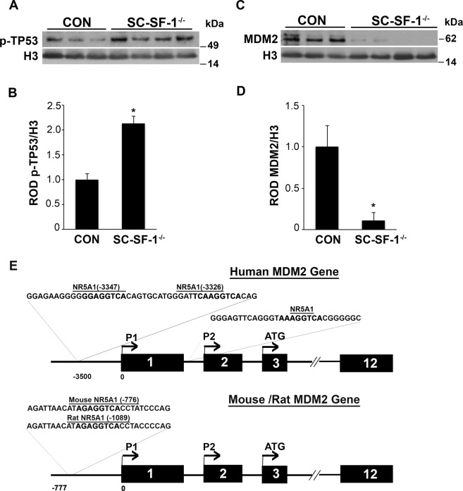
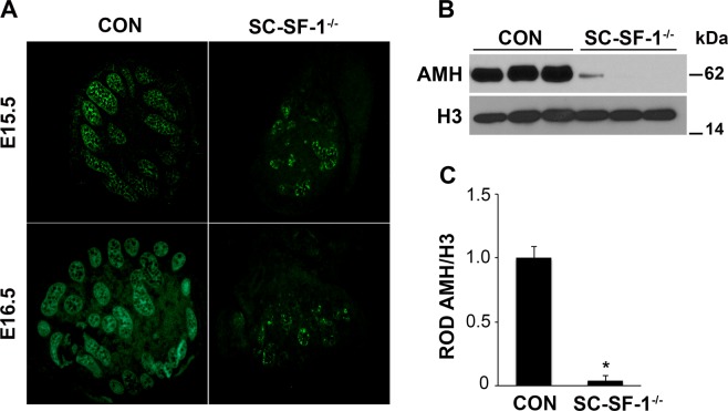
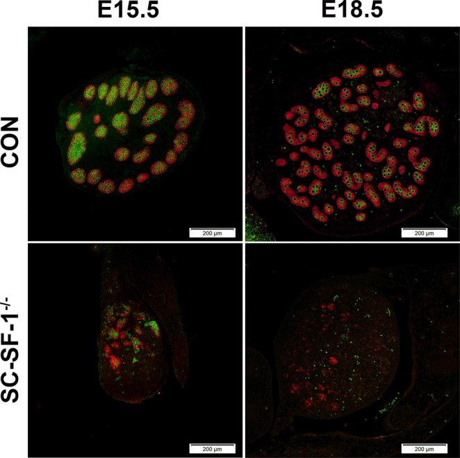
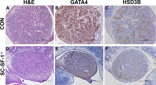
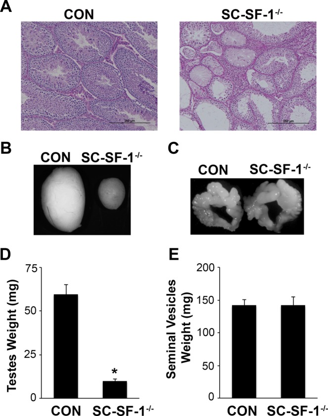
The elevated level of Steroidogenic Factor 1 (Nr5a1, Sf-1) expression in the male gonadal development pathway, post sex determination, implies a vital role in testis gonadal differentiation. In this study we generated Sertoli cell-specific Nr5a1 KO mice (SC-SF-1-/-) at E14.5, which coincides with testis development post sex determination, using the Amh-Cre mouse model. Analysis of SC-SF-1-/- (Sertoli cell specific Nr5a1 knockout) testes demonstrated apoptosis as early as E15. Further analysis revealed that SC-SF-1-/- gonads displayed lower MDM2 levels resulting in elevated TP53 levels, which we believe may lead to apoptosis of the Sertoli cell population, inferring the possibility that NR5A1 directly regulates MDM2 expression. By E15.5, the Sertoli cell and germ cell population declined in SC-SF-1-/- mice resulting in the disruption of seminiferous cords with limited cord structure remaining at E18.5. Due to the loss of Sertoli and germ cells, the testis weights of SC-SF-1-/- mice at 6-weeks were much reduced; however, SC-SF-1-/- seminal vesicles weights were comparable suggesting intact Leydig cell androgen production. We conclude that NR5A1 regulates the TP53 pathway during development, is essential for fetal Sertoli cell survival and controls the cell cycle of Sertoli cells during differentiation.


Conflict of interest statement

The authors declare no competing interests.

Figures

Figure 1
Figure 1
Reduced expression of SOX9 and loss of proliferation of Sertoli cells in SC-SF-1−/− mice. Immunostaining of SOX9 and BrdU in developing testes of control and SC-SF-1−/− mice at E15.5 (A,B), E16.5 (C,D), E17.5 (E,F) and E18.5 (G,H). SC-SF-1−/− testes demonstrated a marked decline in SOX9 positive cells (B,D,F,H) compared to control testes (scale bar = 200 μm). Pregnant mice were injected with BrdU (50 mg/kg b.wt.) 4 h prior to sacrifice. Reduced BrdU incorporation was observed in SC-SF-1−/− testes (B,D,F,H) compared to controls (A,C,E,G). Proliferating Sertoli cells in the seminiferous tubules were identified with SOX9 and BrdU double positive staining (orange; merge 40X). Reduced proliferating Sertoli cells were observed in SC-SF-1−/− testes through E15.5 to E18.5 (IL) compared to their respective controls. (M) SOX9 protein levels were analyzed by western blotting in whole tissue extracts of control and SC-SF-1−/− mice at E15.5. Histone H3 served as a protein loading control. (N) Quantification of western blots demonstrated marked decline in SOX9 expression in SC-SF-1−/− testes compared to control testes. *p < 0.001 by two-tailed Student’s unpaired t test. CON = control.
Figure 2
Figure 2
Apoptosis in Sertoli cell specific NR5A1 knockout testes. TUNEL assay was performed with testes at E15 and E15.5 from control and SC-SF-1−/− embryos to detect apoptotic cells. TUNEL-positive cells were detected using confocal fluorescence microscopy and images were taken at 10X magnification (scale bar = 200 μm). Apoptotic cells were observed in the SC-SF-1−/− testes, but not in the control mice. CON = control. The nuclei are counterstained with DAPI (blue).
Figure 3
Figure 3
Expression of phospho-TP53 (p-TP53) and MDM2 in SC-SF-1−/− mice. The activation of TP53 was investigated in testis of SC-SF-1−/− mice. (A) Western blot and (B) densitometric analysis of p-TP53 (Ser15) in whole tissue extracts of control and SC-SF-1−/− mice at E15. The expression of p-TP53 levels was higher in SC-SF-1−/− testes. (C) MDM2 protein levels were analyzed by western blotting. (D) Densitometric analysis of MDM2 showing a decrease in the protein level. The protein levels were normalized and plotted against Histone H3. *p < 0.01 by two-tailed Student’s unpaired t test. (E) Sequence analyses of 5′ UTR of MDM2 gene in human, rat and mouse species. Human MDM2 gene displaying multiple SF-1 response elements in the P1 (5′ to exon1) and P2 (in intron1) promoter region. Rat and mouse Mdm2 gene have an NR5A1 response element in a highly conserved region in the P1 promoter at position −1089 and −776 respectively upstream of transcription start site. NR5A1 response element sequence is depicted in bold. CON = control.
Figure 4
Figure 4
Loss of NR5A1 in Sertoli cells decreases AMH expression. (A) Immunostaining of AMH in developing testes of control and SC-SF-1−/− mice. Dissected testes from E15.5 and E16.5 demonstrated a marked decline in AMH expression as well as a loss of seminiferous cords in the SC-SF-1−/− embryos. (B) AMH protein levels were analyzed by western blotting in whole tissue extracts of control and SC-SF-1−/− mice at E15.5. Histone H3 served as a protein loading control. (C) Quantification of western blots demonstrated marked decline in AMH expression in SC-SF-1−/− testes compared to control testes. *p < 0.001 by two-tailed Student’s unpaired t test. CON = control.
Figure 5
Figure 5
Nr5a1 ablation in Sertoli cells led to gradual loss of germ cells. Double immunostaining for AMH and VASA was performed in testes at E15.5 and E18.5 from control and SC-SF-1−/− embryos. Sertoli cells labeled by AMH and germ cells labeled by VASA depict a normal cord-like structure in the control testes. A pronounced loss of AMH and VASA staining was observed in SC-SF-1−/− testes suggesting a marked decline in Sertoli cell and germ cell population (scale bar = 200 μm). CON = control.
Figure 6
Figure 6
Morphological changes and Sertoli-cell-only phenotype at E18.5 in SC-SF-1−/− testes. Histological analysis of the testis stained with hematoxylin and eosin (H&E) in control (A) and SC-SF-1−/− (D) mouse embryos at E18.5. H&E staining displayed loss of seminiferous tubules in SC-SF-1−/− testis. Surviving tubules were mostly Sertoli-cell-only (SCO) lacking germ cells. Immunohistochemical staining of GATA4 in control (B) and SC-SF-1−/− (E) testis. GATA4 staining shows the loss of cord formation in SC-SF-1−/− testes. Immunohistochemistry of HSD3B (Leydig cell marker) in testes from control (C) and SC-SF-1−/− (F) mice at E18.5. The Leydig cells were found to express HSD3B in SC-SF-1−/− testes suggesting that loss of Sertoli cells did not affect Leydig cell function (scale bar = 250 μm). CON = control.
Figure 7
Figure 7
NR5A1 depletion in Sertoli cells impeded testes development. (A) Histological analysis of testis sections stained with Periodic Acid-Schiff in control and SC-SF-1−/− mice at 6 weeks of age (scale bar = 250 μm). PAS stain showing abnormal phenotype of the seminiferous tubules including vacuolization and visibly large areas of interstitial cells in SC-SF-1−/− testis. Majority of these seminiferous tubules were devoid of germ cells, forming a Sertoli-cell-only phenotype. Stereomicroscopy images of testes (B) and seminal vesicle (C) from control and SC-SF-1−/− mice at 6 weeks. Average weight of the testes (D) and seminal vesicles (E) of control and SC-SF-1−/− mice. The testes were smaller in SC-SF-1−/− mice, while seminal vesicles were similar in size compared to those of control mice. Values are expressed as the mean ± standard error of the mean (SEM) from 5 mice. *p < 0.001 by two-tailed Student’s unpaired t test. CON = control.

References

    1. Morohashi K, Honda S, Inomata Y, Handa H, Omura T. A common trans-acting factor, Ad4-binding protein, to the promoters of steroidogenic P-450s. J Biol Chem. 1992;267:17913–17919. - PubMed
    1. Rice DA, Mouw AR, Bogerd AM, Parker KL. A shared promoter element regulates the expression of three steroidogenic enzymes. Mol Endocrinol. 1991;5:1552–1561. doi: 10.1210/mend-5-10-1552. - DOI - PubMed
    1. Sadovsky Y, et al. Mice deficient in the orphan receptor steroidogenic factor 1 lack adrenal glands and gonads but express P450 side-chain-cleavage enzyme in the placenta and have normal embryonic serum levels of corticosteroids. Proc Natl Acad Sci USA. 1995;92:10939–10943. doi: 10.1073/pnas.92.24.10939. - DOI - PMC - PubMed
    1. Luo X, Ikeda Y, Parker KL. A cell-specific nuclear receptor is essential for adrenal and gonadal development and sexual differentiation. Cell. 1994;77:481–490. doi: 10.1016/0092-8674(94)90211-9. - DOI - PubMed
    1. Zhao L, et al. Tissue-specific knockouts of steroidogenic factor 1. Molecular and cellular endocrinology. 2004;215:89–94. doi: 10.1016/j.mce.2003.11.009. - DOI - PubMed

Publication types

MeSH terms