. 2019 Mar 14;9(1):4489.
doi: 10.1038/s41598-019-41119-y.

Ultra-fast and automated immunohistofluorescent multistaining using a microfluidic tissue processor

Affiliations

Ultra-fast and automated immunohistofluorescent multistaining using a microfluidic tissue processor

Giulia Cappi et al. Sci Rep. .

Abstract

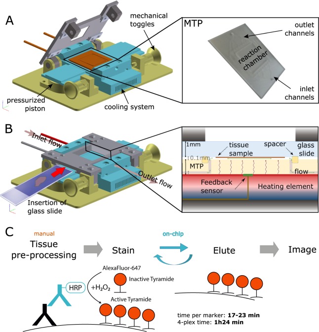
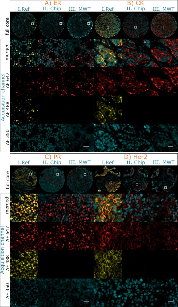
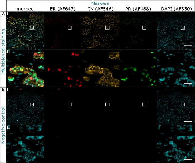
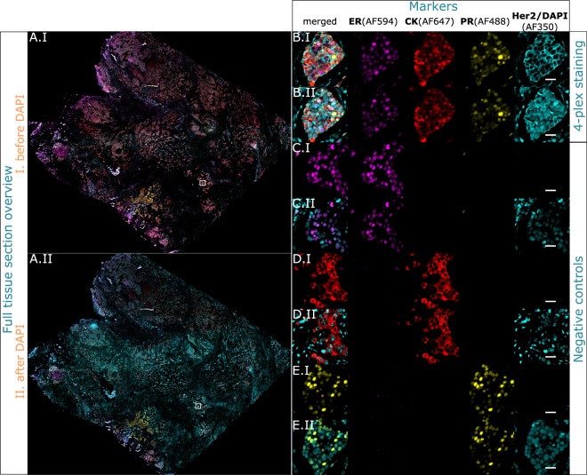
Multistaining of a tissue section targeting multiple markers allows to reveal complex interplays in a tumor environment. However, the resource-intensive and impractically long nature of iterative multiplexed immunostainings prohibits its practical implementation in daily routine, even when using work-flow automation systems. Here, we report a fully automated and ultra-fast multistaining using a microfluidic tissue processor (MTP) in as short as 20 minutes per marker, by immunofluorescent staining employing commercially available tyramide signal amplification polymer precipitation by horse-radish peroxidase (HRP) activation. The reported duration includes (i) 15 minutes for the entire fluidic exchange and reagent incubation necessary for the immunostaining and (ii) 5 minutes for the heat-induced removal of the applied antibodies. Using the automated MTP, we demonstrated a 4-plex automated multistaining with clinically relevant biomarkers within 84 minutes, showing perfect agreement with the state-of-the-art microwave treatment antibody removal. The presented HRP-based method is in principle extendable to multistaining by both tyramides accommodating higher number of fluorescent channels and multi-color chromogenic staining. We anticipate that our automated multi-staining with a turn-around time shorter than existing monoplex immunohistochemistry methods has the potential to enable multistaining in routine without disturbing the current laboratory workflow, opening perspectives for implementation of -omics approaches in tissue diagnostics.


Conflict of interest statement

All the authors are employed by Lunaphore Technologies SA, which filed a patent application covering a portion of the subject matter of this manuscript and which is commercializing the MTP-based technology. D.G.D. and A.T.C. have equity interest in Lunaphore Technologies SA. G.C. has stock options in Lunaphore Technologies SA.

Figures

Figure 1
Figure 1
Microfluidic multiplexing working principle. (A) Sketch of the open stainer showing the position of the MTP inside it. Two mechanical toggles allow for easy opening and closing of the platform. Inset: picture of the MTP. The inset shows the details of the microfluidic inlet and outlet channels. (B) Schematics of the closed stainer and loading of the microscope slide. The slide is inserted laterally such that the tissue is positioned above the reaction chamber. Underneath, a pressurized piston exerts the necessary force to seal the chamber for fast fluid exchange. The inset shows a cut view of the reaction chamber, where the microscope slide hosting the tissue section is clamped together with the MTP chip. The MTP is supported by the heating element, electronically controlled to provide or remove heat from its surface. Cooling grooves facilitate heat dissipation. (C) Work flow of the staining protocol based on the TSA detection system. (i) Tissue pre-processing: slides are manually dehydrated, deparaffinized, re-hydrated and processed for heat-induced epitope retrieval (HIER); (ii) On-chip staining and antibody removal cycles take from 17 to 23 minutes per marker, reaching a total time of 1 h 24 min for a 4-plex staining. The steps performed on-chip are detailed in Table 1; (iii) Slides are finally removed from the stainer, coverslipped and scanned using a multi-spectral epifluorescence microscope. Mechanical stainer designed by Marco Ammann, Lunaphore Technologies SA.
Figure 2
Figure 2
Characterization of the antibody removal efficiency in a 2-staining-cycle protocol. On-chip antibody removal efficiency is demonstrated for ER (A), CK (B), PR (C) and Her2 (D) and compared to the standard MWT. The characterization includes (i) a first detection using TSA-AF647 (Ab-I/Ab-II/TSA-AF647), (ii) heat-induced antibody removal, and (iii) staining with TSA-AF488 to detect the remaining primary and secondary antibodies (PBS/Ab-II/TSA-AF488). The figure shows the image acquisition in the AF647, AF488 and AF350 fluorescent channels, corresponding to a first staining cycle, a second staining cycle and the DAPI counterstain, respectively. (I. Ref) Experiment reference slide with no antibody removal step. All the markers are detected in both AF647 and AF488 channels. (II. Chip) On-chip antibody removal method: the markers are detected only in the AF647channel, while no fluorescent signal is detected in the AF488 channel. (III. MWT) Manual MWT: similarly, the markers are detected only on the AF647 channel. On-chip and MWT methods show equivalent outcome with respect to the evaluation criteria (see main text), however, fully automated on-chip antibody removal in 5 minutes eliminates the need of manual MWT treatment requiring 35 minutes. All the samples have been imaged with the same exposure settings and are visualized with the same parameters. Scale bar: 25 μm.
Figure 3
Figure 3
3-plex staining with primary antibodies originating from the same species obtained with the on-chip antibody removal method. (A) Multiplexed colocalized staining of ER, CK and PR. (B) Negative staining where the primary antibodies were replaced by washing buffer. All the markers were specifically stained and detected in their corresponding detection channel: AF647 for ER, AF546 for CK, AF488 for PR and AF350 for DAPI. No crosstalk between channels of subsequent stainings indicates the efficient removal of antibodies originating from the same species and no damage to the epitopes. For every panel: row I is the overview image, scale bar 500 μm; row II is a zoom-in where the 3 markers are co-expressed, scale bar 25 μm.
Figure 4
Figure 4
Multiplexed colocalized staining on-chip of ER (AF594), CK (AF647), PR (AF488) and Her2 (AF350) in 84 minutes. Full-tissue staining on a breast carcinoma sample for ER, CK, PR and Her2 is achieved in 84 minutes, excluding slide pre-processing. All the markers are specifically detected. Her2 and DAPI are visualized in the same channel (AF350) by imaging before and after counterstaining. (A) Overview image of the whole tissue section. (BE) Zoom-in area where the 4 markers are co-expressed, visualized in the 4 acquisition channels. (B) 4-plex protocol. (C) Negative control where only ER is stained to be used as a focus reference. (D) Negative control where only CK is stained to be used as a focus reference. (E) Negative control where only PR is stained to be used as a focus reference. For every sequence panel: (I) is the first image acquisition without nuclei counterstain, (II) is the second acquisition after the nuclei were counterstained with DAPI. Scale bar 25 μm.
Figure 5
Figure 5
Colocalized 4-plex staining of ER (AF647), CK (AF546), PR (AF488) and Her2 (AF350) applied to the breast TMA. 4-plex staining performed on the same cores used for the antibody removal characterization in Fig. 2. Specific staining is observed for each marker. The left column shows the overview of the TMA cores, scale bar 500 μm. Columns 2–6: (A) Core employed for the antibody removal characterization of ER (Fig. 2A), expressing also PR and CK staining and negative to Her2. (B) Core employed for the antibody removal characterization of CK (Fig. 2B), expressing also ER and PR, and negative to Her2. (C) Core employed for the antibody removal characterization of PR (Fig. 2C), expressing also CK and negative to ER and Her2. (D) Core employed for the antibody removal characterization of Her2 (Fig. 2D), expressing also CK and negative to ER and PR. Scale bar 25 μm.

References

    1. Jürgensmeier, J. M., Eder, J. P. & Herbst, R. S. New Strategies in Personalized Medicine for Solid Tumors: Molecular Markers and Clinical Trial Designs. Clinical cancer research: an official journal of the American Association for Cancer Research20, 4425–4435, http://www.ncbi.nlm.nih.gov/pmc/articles/PMC5369358/, 10.1158/1078-0432.CCR-13-0753 (2014). - PMC - PubMed
    1. Hicks DG. T. P. Molecular classifications of breast carcinoma: is immunohistochemistry a viable alternative to other molecular methodologies? Connection. 2009;13:31–34.
    1. Tan, D. & Zander, D. S. Immunohistochemistry for Assessment of Pulmonary and Pleural Neoplasms: A Review and Update. International Journal of Clinical and Experimental Pathology1, 19–31, http://www.ncbi.nlm.nih.gov/pmc/articles/PMC2480532/ (2008). - PMC - PubMed
    1. Jagirdar J. Application of immunohistochemistry to the diagnosis of primary and metastatic carcinoma to the lung. Archives of pathology & laboratory medicine. 2008;132:384–396. doi: 10.1043/1543-2165. - DOI - PubMed
    1. Capelozzi VL. Role of immunohistochemistry in the diagnosis of lung cancer. Jornal brasileiro de pneumologia: publicacao oficial da Sociedade Brasileira de Pneumologia e Tisilogia. 2009;35:375–382. doi: 10.1590/S1806-37132009000400012. - DOI - PubMed

MeSH terms