. 2019 Feb 28:12:49.
doi: 10.3389/fnmol.2019.00049. eCollection 2019.

An Intersectional Approach to Target Neural Circuits With Cell- and Projection-Type Specificity: Validation in the Mesolimbic Dopamine System

Affiliations

An Intersectional Approach to Target Neural Circuits With Cell- and Projection-Type Specificity: Validation in the Mesolimbic Dopamine System

Nefeli Kakava-Georgiadou et al. Front Mol Neurosci. .

Abstract

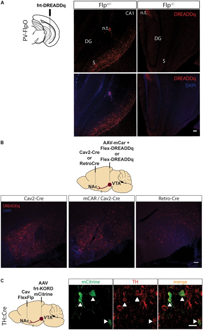
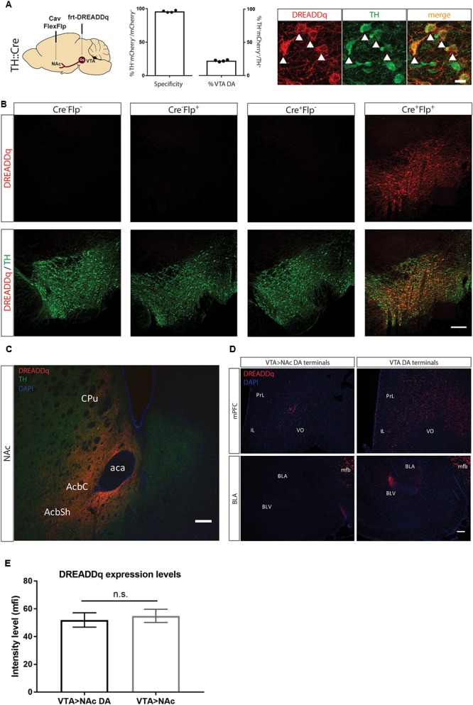
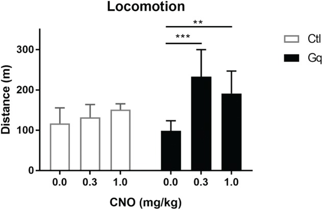
Development of tools to manipulate activity of specific neurons is important for dissecting the function of neural circuits. Viral vectors and conditional transgenic animal lines that target recombinases to specific cells facilitate the successful manipulation and recording of specific subsets of neurons. So far, it has been possible to target neuronal subtypes within a certain brain region based on transcriptional control regions from a gene selectively expressed in those cells or based upon its projections. Nevertheless, there are only a few tools available that combine this and target a neuronal subtype within a projection. We tested a viral vector system, consisting of a canine adenovirus type 2 expressing a Cre-dependent Flp recombinase (CavFlexFlp) and an adeno-associated viral (AAV) vector expressing a Flp-dependent cDNA, which targets neurons in a subtype- and projection-specific manner. As proof of principle we targeted expression of a Designer Receptor Exclusively Activated by Designer Drugs (DREADD) to the dopamine neurons of the mesolimbic projection, which allows the transient activation of neurons by the ligand Clozapine-N-Oxide (CNO). We validated that the system specifically targets dopamine neurons and that chemogenetic activation of these neurons induces an increase in locomotor activity. We thus validated a valuable tool that allows in vivo neuronal activation in a projection- and subtype-specific manner.

Keywords: CNO; Cav2; DREADD; Flp; VTA; canine; chemogenetics; dopamine.


Figures

FIGURE 1
FIGURE 1
Pilot testing of viral vectors. (A) PV-FlpO+/- and -/- mice were injected in the ventral hippocampus with frt-DREADDq. Immunohistochemistry against mCherry (fused to DREADDq) showed expression of DREADDq in sections from ventral hippocampus of PV-FlpO+/- but not of PV-FlpO-/- littermates. (B) Rats were injected in the NAc and VTA with a combination of Cav2-Cre and lox-DREADDq (Cav2-Cre) or Cav2-Cre and lox-DREADDq with AAV-mCAR (mCAR/Cav2-Cre) or Retro-Cre and lox-DREADDq (Retro-Cre) respectively. Immunohistochemical detection of mCherry (fused to DREADDq) showed no difference in expression levels between Cav2-Cre and mCAR/Cav2-Cre, whereas Retro-Cre brought lower levels of mCherry expression. (C) TH::Cre rats were injected with CavFlexFlp and frt-KORD-mCitrine in the NAc and VTA, respectively, so that VTA>NAc dopamine neurons would be targeted. Immunohistochemistry against mCitrine (green) and TH (red) showed co-localization of the mCitrine+ with TH+ neurons (white arrows). n.t., needle tract; CA1, field CA1 of the hippocampus; DG, Dentate Gyrus; S, Subicculum.
FIGURE 2
FIGURE 2
Targeting VTA>NAc dopamine neurons with DREADDq. (A) TH::Cre+/- rats were injected in the NAc and VTA with CavFlexFlp and frt-DREADDq, respectively. Co-staining for mCherry (fused to DREADDq, red) and TH (green), showed co-localization (white arrows), with 22,2% ± 0,6 of the total VTA TH+ neurons targeted (% mCherry+TH+ / total TH+) and specificity of 95,9% ± 0,8 (% mCherry+TH+ / total mCherry+). (B) TH::Cre-/- rats were injected with frt-DREADDq in the VTA (Cre-Flp-) or with CavFlexFlp in the NAc and frt-DREADDq in the VTA (Cre-Flp+); TH:Cre+/- rats were injected with frt-DREADDq in the VTA (Cre+Flp-) or with CavFlexFlp in the NAc and frt-DREADDq in the VTA (Cre+Flp+); Immunohistochemical detection of mCherry (fused to DREADDq) showed no expression of DREADDq in Cre-Flp-, Cre-Flp+ and Cre+Flp- rats, whereas in Cre+Flp+ rats DREADDDq was expressed in the VTA. (C) Fibers positive for mCherry (red) were detected in the NAc shell and core, where TH+ fibers are also detected (green) in TH::Cre+/- rats injected in the NAc and VTA with CavFlexFlp and frt-DREADDq, respectively. (D) In TH::Cre+/- rats injected in the VTA with lox-DREADDq (VTA DA) fibers positive for mCherry (fused to DREADDq) were detected in the mPFC and the BLA (panels on the right), whereas in TH::Cre+/- rats injected in the NAc and VTA with CavFlexFlp and frt-DREADDq, respectively few or no mCherry+ fibers were detected in the mPFC and the BLA, respectively (panels on the left). (E) Expression levels of DREADDq were measured by quantification of mCherry intensity (as mean fluorescent intensity – mfi) in TH::Cre+/- rats injected in the NAc and VTA with CavFlexFlp and frt-DREADDq, respectively (VTA>NAc DA) and TH::Cre-/- rats injected in the NAc and VTA with Cav2-Cre and lox-DREADDq, respectively (VTA>NAc). No difference was found between VTA>NAc DA and VTA>NAc targeting (unpaired t-test; t(25) = 0,4093, p = 0.6858); Data represented as mean ± SEM; ns, not significant; Cpu, Caudate putamen; AcbC, Accumbens nucleus, Core; AcbSh, Accumbens nucleus, Shell; aca, anterior commissure; PrL, Prelimbic cortex; IL, Infralimbic cortex; VO, Ventral Orbital cortex; BLA, Basolateral Amygdaloid nucleus, anterior; BLV, Basolateral Amygdaloid nucleus, ventral; mfb, medial forebrain bundle.
FIGURE 3
FIGURE 3
Activation of VTA>NAc DA neurons increases locomotor activity. Gq rats were TH::Cre+/- rats injected in the NAc and VTA with CavFlexFlp and frt-DREADDq, respectively (n = 13); Ctl rats were either TH::Cre+/- rats injected in the NAc and VTA with CavFlexFlp and frt-mCherry, respectively, or TH::Cre-/- rats injected in the NAc and VTA with CavFlexFlp and frt-DREADDq, respectively (total n = 9); CNO administration did not increase locomotor activity in Ctl rats (Friedman test; χ2(9) = 1.556, p = 0.5690), whereas CNO significantly increased locomotor activity in Gq rats (Friedman test; χ2(13) = 0.333, p = 0.0002; Dunn’s multiple comparisons post hoc test: Saline vs. CNO 0.3 mg/kg p = 0.0004; Saline vs. CNO 1.0 mg/kg p = 0.0017); Data represented as median with interquartile range; ∗∗P < 0.01, ∗∗∗P < 0.001.

Similar articles

Cited by

References

    1. Aransay A., Rodríguez-López C., García-Amado M., Clascá F., Prensa L. (2015). Long-range projection neurons of the mouse ventral tegmental area: a single-cell axon tracing analysis. Front. Neuroanat. 9:59. 10.3389/fnana.2015.00059 - DOI - PMC - PubMed
    1. Atasoy D., Betley J. N., Su H. H., Sternson S. M. (2012). Deconstruction of a neural circuit for hunger. Nature 488 172–177. 10.1038/nature11270 - DOI - PMC - PubMed
    1. Augur I. F., Wyckoff A. R., Aston-Jones G., Kalivas P. W., Peters J. (2016). Chemogenetic activation of an extinction neural circuit reduces cue-induced reinstatement of cocaine seeking. J. Neurosci. 36 10174–10180. 10.1523/JNEUROSCI.0773-16.2016 - DOI - PMC - PubMed
    1. Backer M. W. A. (2010). Optimization of Viral Vector Technology to Study Gene Function in the Hypothalamus. Available at: http://dspace.library.uu.nl/handle/1874/44371 [accessed April 21 2014].
    1. Betley J. N., Sternson S. M. (2011). Adeno-Associated viral vectors for mapping, monitoring, and manipulating neural circuits. Hum. Gene Ther. 22 669–677. 10.1089/hum.2010.204 - DOI - PMC - PubMed