. 2019 Feb 28:11:44.
doi: 10.3389/fnagi.2019.00044. eCollection 2019.

Dl-3- n-Butylphthalide Exerts Dopaminergic Neuroprotection Through Inhibition of Neuroinflammation

Affiliations

Dl-3- n-Butylphthalide Exerts Dopaminergic Neuroprotection Through Inhibition of Neuroinflammation

Yajing Chen et al. Front Aging Neurosci. .

Erratum in

Abstract

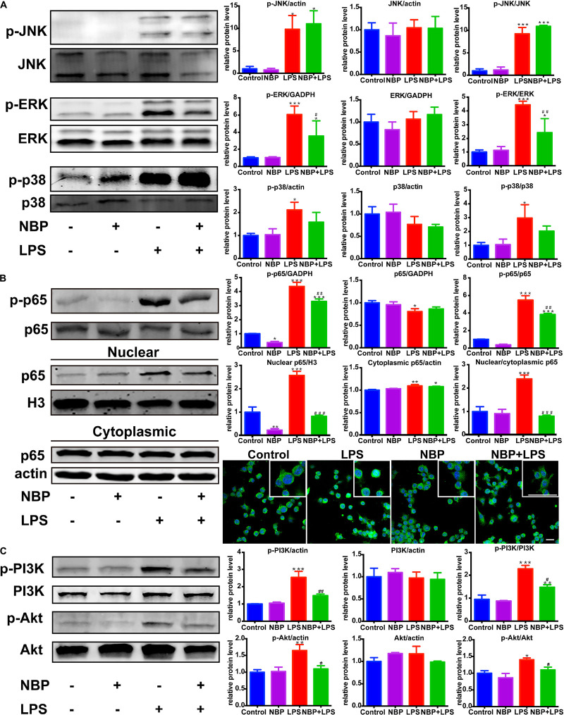
Microglia-mediated neuroinflammation contributes to multiple neurodegenerative disorders, including PD. Therefore, the regulation of microglial activation probably has the therapeutic potential. This study is aimed to determine whether NBP could suppress microglial activation and protect dopaminergic neurons from excessive neuroinflammation. In the present study, MPTP-induced PD model was established to explore the neuroprotective and anti-inflammatory effect of NBP. We assessed motor deficits, dopaminergic neurodegeneration and microglial activation in PD mice. In vitro, the anti-inflammatory activity of NBP was confirmed by cell viability assay of SH-SY5Y cells after being treated with conditioned medium from LPS-stimulated BV-2 cells and from 1-Methyl-4-phenylpyridinium iodide (MPP+)-stimulated BV-2 cells. The expression of pro-inflammatory molecules was determined by RT-PCR, Western Blot and ELISA assay. The generation of NO and ROS were also assessed. The involvement of signaling pathways such as MAPK, NF-κB, and PI3k/Akt were further investigated by Western Blot and immunofluorescence assay. The neuroprotective effect of NBP was demonstrated in vivo as shown by the improvement of dopaminergic neurodegeneration, motor deficits and microglial activation in MPTP-induced mouse model of PD. The expression of pro-inflammatory mediators was also reduced by NBP administration. In vitro, NBP also protected dopaminergic neurons from neurotoxicity induced by activated microglia. NBP pretreatment not only reduced pro-inflammatory molecules, but also suppressed NO release and ROS generation in BV-2 cells. Further mechanism research suggested that the inactivation of MAPK, NF-κB and PI3K/Akt may involve in anti-neuroinflammation role of NBP. In conclusion, our results revealed that NBP exerted dopaminergic neuroprotection through inhibition of microglia-mediated neuroinflammation, suggesting the promising therapeutic effect of NBP for PD.

Keywords: MAPK; NF-κB; Parkinson’s disease; dl-3-n-butylphthalide; microglia; neuroinflammation.


Figures

Figure 1
Figure 1
NBP improved MPTP-induced motor deficits. (A) The Experimental procedure and drug administration scheme. (B,C) Total distance (B) and mean velocity (C) of MPTP-treated mice in the open field test. (D,E) Total time (D) and turn time (E) spent in the pole test. (F) Score of hanging wire test. (G) Latency to fall in the rotarod test. All data are presented as means ± SEM (n = 10). p < 0.05, ∗∗p < 0.01, ∗∗∗p < 0.001, compared with the Control group; #p < 0.05, ###p < 0.001, compared with the MPTP group.
Figure 2
Figure 2
NBP ameliorated MPTP-induced nigrostriatal dopaminergic injury. (A) Immunohistochemical staining showing TH positive cells in the SN (scale bar: 0.2 mm). Stereological counting of TH positive cells is shown in the right panel (n = 4). (B) Immunohistochemical staining showing striatal TH positive nerve fibers (scale bar: 0.2 mm). Densitometric analysis of the relative optical density of TH staining is shown in the right panel (n = 4). (C) Western Blot analysis of striatal TH protein (n = 4). Protein band intensity is normalized to actin and is expressed as fold difference relative to the Control group. (D) HPLC assays of DA, DOPAC, HVA, 5-HT and 5-HIAA (n = 5). All data are presented as means ± SEM. p < 0.05, ∗∗p < 0.01, ∗∗∗p < 0.001, compared with the Control group; #p < 0.05, ##p < 0.01, ###p < 0.001, compared with the MPTP group.
Figure 3
Figure 3
NBP suppressed MPTP-induced activation of microglia in the striatum and SN. (A) Immunohistochemical staining showing Iba-1 positive cells in the striatum (scale bar: 0.1 mm). Counting of Iba-1 positive cells in the striatum is shown in the right panel (n = 4). (B) Double immunofluorescence staining of TH (red) and Iba-1 (green) in the SN (scale bar: 50 μm). Relative immunoreactivity intensity of Iba-1 is shown in the right panel (n = 3). All data are presented as means ± SEM. ∗∗∗p < 0.001, compared with the Control group; ###p < 0.001, compared with the MPTP group.
Figure 4
Figure 4
NBP reduced the expression of inflammatory mediators and inhibited the MAPK-ERK pathway in MPTP-treated mice. (A) Western Blot analysis of striatal IL-1β and COX-2 protein. (B) Double immunofluorescence staining of iNOS (red) and Iba-1 (green) in the SN (scale bar: 25 μm). (C) Western Blot assay for MAPKs pathway. All data are presented as means ± SEM (n = 3–4). p < 0.05, ∗∗p < 0.01, compared with the Control group; #p < 0.05, ##p < 0.01, compared with the MPTP group.
Figure 5
Figure 5
NBP protected dopaminergic neurons from neurotoxicity induced by microglial activation. SH-SY5Y cells were incubated for 24 h with conditioned medium derived from cultures of BV-2 cells. Before collecting culture media, BV-2 cells were pretreated with NBP (0 or 100 μM) for 1 h and incubated with LPS (0 or 100 ng/ml) or MPP+ (0 or 500 μM) for 24 h. (A) Cell viability was measured with the CCK8 assay (n = 5). All data are presented as means ± SEM. ∗∗p < 0.01, ∗∗∗p < 0.001, compared with the Control group; #p < 0.05, ##p < 0.01, ###p < 0.001, compared with the LPS group or the MPP+ group. (B) The apoptosis of SH-SY5Y was evaluated by immunofluorescence detection of cleaved caspase-3 (green) and cell nuclei was stained with DAPI (blue) (scale bar: 50 μm). (C) The cell death of SH-SY5Y was evaluated by immunofluorescence detection of PI (red) (scale bar: 50 μm).
Figure 6
Figure 6
NBP reduced pro-inflammatory molecules expression in LPS-stimulated BV-2 cells. BV-2 cells were pretreated with NBP (0 or 100 μM) for 1 h followed by LPS (0 or 100 ng/ml) for 6 h and total RNA was isolated. To measure cellular protein expression or cytokines level in supernatants, time for LPS treatment was 24 h. (A) The mRNA expression of IL-1β, IL-6, TNF-a, iNOS and COX-2 was analyzed by RT-PCR and normalized to that of β-actin. (B) The protein level of TNF-α, IL-1β, iNOS and COX2 was analyzed by Western Blot. (C) The level of TNF-α and IL-1β in supernatants was assayed by ELISA kit. All data are presented as means ± SEM (n = 3). p < 0.05, ∗∗p < 0.01, ∗∗∗p < 0.001, compared with the Control group; #p < 0.05, ##p < 0.01, ###p < 0.001, compared with the LPS group.
Figure 7
Figure 7
NBP suppressed NO release and ROS generation in LPS-stimulated BV-2 cells. BV-2 cells were pretreated with NBP (0 or 100 μM) for 1 h and followed by LPS (0 or 100 ng/ml) for 24 h. (A) NO levels in culture supernatant were measured by Griess method (n = 3). (B) ROS production in BV-2 was measured using fluorescence probe DCFH-DA and the fluorescence intensity was determined at 485 nm excitation and 535 nm emission (n = 5). (C) Immunofluorescence detection of DCFH-DA (green) (scale bar: 0.1 mm). All data are presented as means ± SEM (n = 3). ∗∗∗p < 0.001, compared with the Control group; ###p < 0.001, compared with the LPS group.
Figure 8
Figure 8
NBP inhibited the activation of ERK, NF-κB and PI3K/Akt pathways in LPS-stimulated BV-2 cells. (A) Western Blot assay for MAPK expression. The expression of total JNK/p38/ERK as well as p-JNK/p-p38/p-ERK were analyzed. (B) Western Blot assay for NF-κB expression in whole-cell, nuclear and cytoplasmic extracts. The nuclear translocation of p65 was also evaluated by immunofluorescence detection of p65 (green) and cell nuclei was stained with DAPI (blue) (scale bar: 50 μm). (C) Western Blot assay for PI3K/Akt expression. The expression of total PI3K/Akt as well as p-PI3K/p-Akt were analyzed. All data are presented as means ± SEM (n = 3). p < 0.05, ∗∗p < 0.01, ∗∗∗p < 0.001, compared with the Control group; #p < 0.05, ##p < 0.01, ###p < 0.001, compared with the LPS group.
Figure 9
Figure 9
NBP reduced IL-1β level and inhibited the activation of JNK and NF-κB pathways in MPP+-stimulated BV-2 cells. BV-2 cells were pretreated with NBP (0 or 100 μM) for 1 h and followed by MPP+ (0 or 500 μM) for 24 h. (A) The protein level of IL-1β was analyzed by Western Blot. (B) Western Blot assay for MAPK and NF-κB expression. All data are presented as means ± SEM (n = 3). ∗∗p < 0.01, ∗∗∗p < 0.001, compared with the Control group; #p < 0.05, ##p < 0.01, ###p < 0.001, compared with the MPP+ group.
Figure 10
Figure 10
Schematic illustration demonstrates that NBP protects dopaminergic neurons from inflammation via inhibition of MAPK, NF-κB and PI3K/Akt pathways in microglia.

References

    1. Asakawa T., Fang H., Sugiyama K., Nozaki T., Hong Z., Yang Y., et al. (2016). Animal behavioral assessments in current research of parkinson’s disease. Neurosci. Biobehav. Rev. 65 63–94. 10.1016/j.neubiorev.2016.03.016 - DOI - PubMed
    1. Athauda D., Foltynie T. (2015). The ongoing pursuit of neuroprotective therapies in parkinson disease. Nat. Rev. Neurol. 11 25–40. 10.1038/nrneurol.2014.226 - DOI - PubMed
    1. Bhatia H. S., Baron J., Hagl S., Eckert G. P., Fiebich B. L. (2016). Rice bran derivatives alleviate microglia activation: possible involvement of MAPK pathway. J. Neuroinflamm. 13:148. 10.1186/s12974-016-0615-6 - DOI - PMC - PubMed
    1. Cao J. P., Wang H. J., Yu J. K., Liu H. M., Gao D. S. (2008). The involvement of NF-kappaB p65/p52 in the effects of GDNF on DA neurons in early PD rats. Brain Res. Bull. 76 505–511. 10.1016/j.brainresbull.2008.03.007 - DOI - PubMed
    1. Chen Y., Jiang M., Li L., Ye M., Yu M., Zhang L., et al. (2018). DL3nbutylphthalide reduces microglial activation in lipopolysaccharideinduced parkinson’s disease model mice. Mol. Med. Rep. 17 3884–3890. 10.3892/mmr.2017.8332 - DOI - PubMed

LinkOut - more resources