. 2019 Feb 28:10:122.
doi: 10.3389/fphys.2019.00122. eCollection 2019.

Biochemical and Functional Characterization of Glycoside Hydrolase Family 16 Genes in Aedes aegypti Larvae: Identification of the Major Digestive β-1,3-Glucanase

Affiliations

Biochemical and Functional Characterization of Glycoside Hydrolase Family 16 Genes in Aedes aegypti Larvae: Identification of the Major Digestive β-1,3-Glucanase

Raquel Santos Souza et al. Front Physiol. .

Abstract

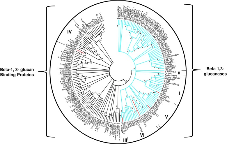
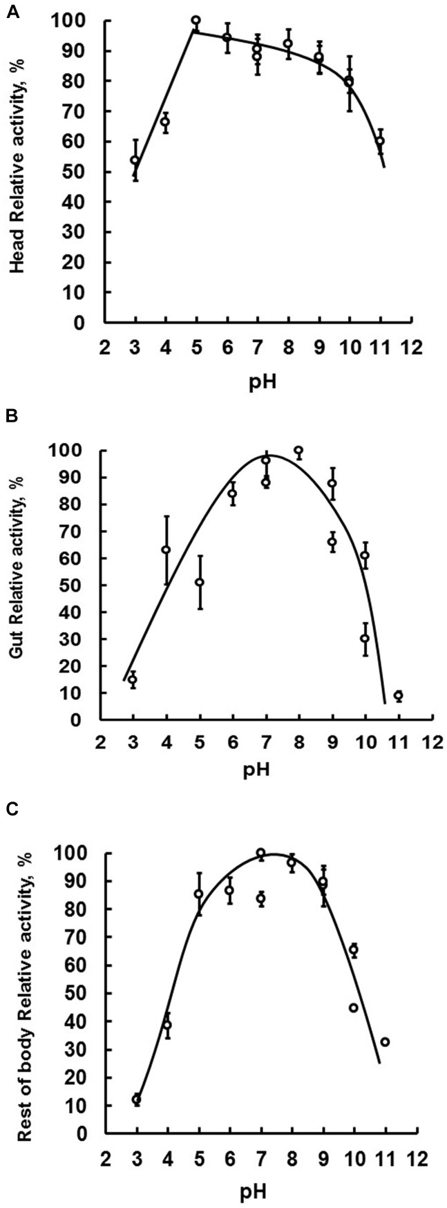
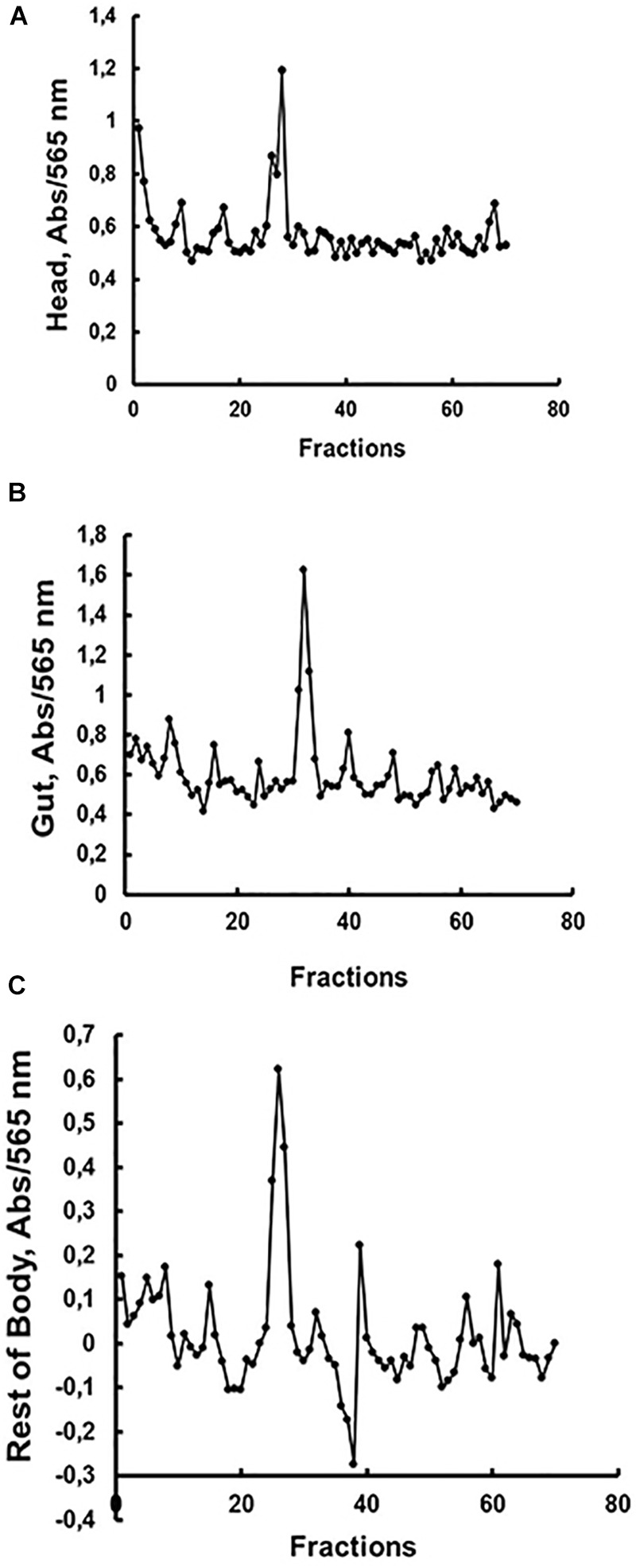
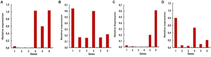
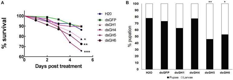
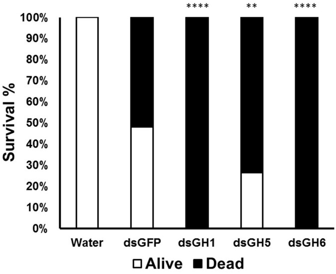
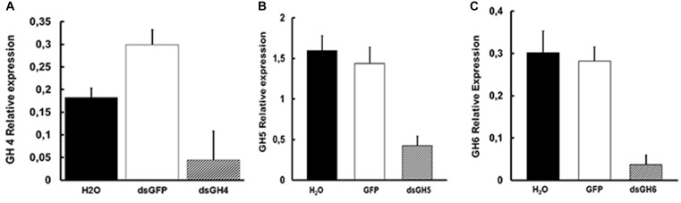
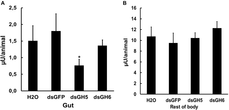
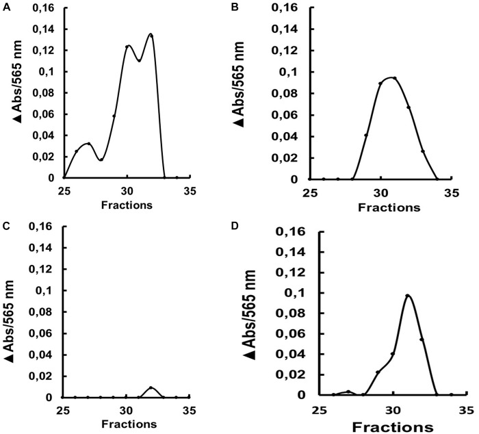
Insect β-1,3-glucanases belong to Glycoside Hydrolase Family 16 (GHF16) and are involved in digestion of detritus and plant hemicellulose. In this work, we investigated the role of GHF16 genes in Aedes aegypti larvae, due to their detritivore diet. Aedes aegypti genome has six genes belonging to GHF16 (Aae GH16.1 - Aae GH16.6), containing two to six exons. Sequence analysis suggests that five of these GHF16 sequences (Aae GH16.1, 2, 3, 5, and 6) contain the conserved catalytic residues of this family and correspond to glucanases. All genomes of Nematocera analyzed showed putative gene duplications corresponding to these sequences. Aae GH16.4 has no conserved catalytic residues and is probably a β-1,3-glucan binding protein involved in the activation of innate immune responses. Additionally, Ae. aegypti larvae contain significant β-1,3-glucanase activities in the head, gut and rest of body. These activities have optimum pH about 5-6 and molecular masses between 41 and 150 kDa. All GHF16 genes above showed different levels of expression in the larval head, gut or rest of the body. Knock-down of AeGH16.5 resulted in survival and pupation rates lower than controls (dsGFP and water treated). However, under stress conditions, severe mortalities were observed in AeGH16.1 and AeGH16.6 knocked-down larvae. Enzymatic assays of β-1,3-glucanase in AeGH16.5 silenced larvae exhibited lower activity in the gut and no change in the rest of the body. Chromatographic activity profiles from gut samples after GH16.5 silencing showed suppression of enzymatic activity, suggesting that this gene codes for the digestive larval β-1,3-glucanase of Ae. aegypti. This gene and enzyme are attractive targets for new control strategies, based on the impairment of normal gut physiology.

Keywords: Aedes aegypti; GHF16; Glycoside Hydrolase Family 16; digestion; immunity; knock-down; β-1,3-glucanase.


Figures

FIGURE 1
FIGURE 1
Cladogram of selected protein sequences of insect β-1,3-glucanases, and β-1,3-glucan binding proteins. Branches are statistically supported by bootstrap analysis (cutoff 70%). The blue branches discriminate the monophyletic sequences of the insects that have the conserved catalytic glutamates; the green branches discriminate the paraphyletic group of sequences that do not have the conserved catalytic residues. The bootstrap values were obtained from the analysis of 10,000 replicates, using the Neighbor-Joining algorithm (MEGA software 5.05). Consensus phylogenetic tree used sequences of: Anopheles gambiae (AGAP002798-PA, AGAP002799-PA, AGAP002796-PA, AGAP006761-PA, AGAP012409-PA), Anopheles christyi (ACHR004102-RA, ACHR005689, ACHR008721-RA, ACHR001881-RA, ACHR009179-RA), Anopheles darlingi (ADAR007290-PA, ADAR007286-PA, ADAR006526-PA, ADAR009199-PA), Anopheles dirus (ADIR003516-RA, ADIR010616-RA, ADIR003518-RA, ADIR003625-RA, ADIR000553-RA) Anopheles epiroticus (AEPI010194-RA, AEPI009256-RA, AEPI005496-RA, AEPI002293-RA), Anopheles funestus (AFUN006014-RA, AFUN009437-RA, AFUN006016-RA, AFUN002755-RA, AFUN004083-RA), Anopheles minimus (AMIN004837-RA, AMIN003902-RA, AMIN003903-RA, AMIN003900-RA, AMIN010081-RA, AMIN008919-RA), Anopheles quadriannulatus (AQUA008516-RA, AQUA009400-RA, AQUA009402-RA, AQUA003848-RA, AQUA014348-RA), Anopheles stephensi (ASTE003966-RA, ASTE009324-RA, ASTE009326-RA, ASTE010371-RA, ASTE004573-RA), Culex quinquefasciatus (XM_001845911.1, XM_001845228.1, XM_001845913.1, XM_001845759.1, JF907421.1, XM_002135149.1, XM_001845915.1, XM_001847484.1, XM_001847484.1, XM_001845910.1, XM_001845757.1, XM_001864211.1, XM_001845229.1), Phlebotomus papatasi (PPATMP000880-PA, PPATMP002587-PA, PPATMP002588-PA, PPATMP010440-PA), Rhodnius prolixus (RPRC011769-PA, RPRC003210-PA, ABU96697.1), Simulium vittatum (EU930267.1), Anopheles arabiensis (ACN38171.1, CAO83421.1), Anopheles bwambae (ABU80038.1), Anopheles melas (ABU80011.1), Anopheles merus (ABU80005.1, AAZ08489.1, AAZ08502.1), Ochlerotatus triseriatus (ACU30929.1), Phlebotomus perniciosus (ADH94599.1). The code of the other sequences can be seen in Supplementary Table S3.
FIGURE 2
FIGURE 2
The tissue-specific optimal enzymatic activity of β-1,3-glucanase. Samples were assayed using laminarin as substrate. Optimal activity was determined by performing enzymatic reactions under a pH range of 3–11. Evaluated tissues were head (A), digestive tract (B), and rest of the body (C) of fourth instar larval Ae. aegypti.
FIGURE 3
FIGURE 3
Activity against laminarin in fractions obtained after gel filtration chromatography (Superdex 200 – AKTA FPLC) of soluble fractions obtained from the (A) head, (B) digestive tract and (C) rest of the body of Ae. aegypti larvae. Elution volumes and molecular masses of protein standards were used to build a calibration curve for calculation of the molecular masses of β-1,3-glucanase activities. Elution volumes (mL)/fraction (number) of the standards were as follows: 18.22/34 (cytochrome C, 12.4 kDa), 16.86/32 (carbonic anhydrase, 29 kDa), 14.87/28 (bovine serum albumin, 66 kDa), 13.49/25 (alcohol dehydrogenase, 150 kDa), 12.77/24 (β-amylase, 200 kDa), and 5.64/9 (blue dextran, 2000 kDa). The elutions (mL/fraction number) of β-1,3-glucanase activities from (A) head, (B) gut, and (C) rest of body were 13.75/26, 16.75/32, and 13.5/25, respectively, resulting in predicted molecular masses of 142, 41, and 150 kDa.
FIGURE 4
FIGURE 4
Relative expressions of the genes encoding GHF16 proteins in fourth instar larvae of Ae. aegypti, measured in series of semi-quantitative RT-PCR reactions with 24 or 27 cycles. Numbers are relative expressions normalized using the gene RP49 as a constitutive control. cDNA samples were prepared as described in “Materials and Methods” section from (A) whole L4 larvae (27 cycles), (B) heads (27 cycles), (C) guts (24 cycles) and (D) rest of bodies (27 cycles). 1–6 correspond to the relative expression levels of AaeGH16.1-6, respectively.
FIGURE 5
FIGURE 5
Ae. aegypti GHF16 genes have distinct physiological roles. (A) Survival curve of Ae. aegypti fourth instar larvae after feeding with dsRNA. dsGH1, dsGH4, dsGH5, dsGH6 correspond to larvae fed with dsRNA coding, respectively, for the genes AaeGH16.1, AaeGH16.4, AaeGH16.5, and AaeGH16.6. GFP and H2O correspond to the control groups fed with dsGFP or water only. (B) Percentage of Ae. aegypti fourth instar larvae and pupae in after treatment with dsRNA and following for 12 days in control diet (cat food). GH1/GH4/GH5/GH6/GFP/H2O correspond to larvae exposed to dsRNA coding for AaeGH16.1, AaeGH16.4, AaeGH16.5, AaeGH16.6, GFP, and water, respectively. Comparison of survival curves vs. the control treated with dsGFP used the Log-rank test (p < 0.05; ∗∗p < 0.01; ∗∗∗p < 0.001) and comparison of groups vs. the control treated with dsGFP used the chi-square test (p < 0.05; ∗∗p < 0.01).
FIGURE 6
FIGURE 6
Mortality and survival of Ae. aegypti fourth instar larvae treated with dsRNA under conditions of high density and food restriction. GH1, GH5, and GH6 correspond to insects treated with dsRNA coding for AaeGH16.1, AaeGH16.5, and AaeGH16.6, respectively. GFP and H2O correspond to controls treated with dsRNA coding for GFP and water, respectively. Comparison of groups vs the control treated with dsGFP used the Fisher’s exact test (∗∗p < 0.01; ∗∗∗∗p < 0.0001).
FIGURE 7
FIGURE 7
Levels of expression of the genes AeGH16.4 (GH4), AeGH16.5 (GH5), and AeGH16.6 (GH6) in Ae. aegypti fourth instar larvae after treatment with dsRNA. (A) Relative expression of the gene AeGH16.4 in whole larvae. (B) Relative expression of the gene AeGH16.5 in the gut. (C) Relative expression of the gene AeGH16.6 in the rest of the body. Expression levels were normalized using the ribosomal RP gene 49 as the constitutive marker. We used 24 cycles for AeGH16.4 and 27 cycles for AeGH16.5 and AeGH16.6 in the RT-PCR reactions. See “Materials and Methods” for details.
FIGURE 8
FIGURE 8
β-1,3-glucanase activity (laminarin as substrate) in the soluble fractions of the digestive tract (A) and rest of the body (B) of fourth instar larvae of Ae. aegypti treated with water (H2O) or dsRNA coding for GFP, AaeGH16.5 (dsGH5), and AaeGH16.6 (dsGH6). Comparison of groups vs. the control treated with dsGFP used the t-test (p < 0.05).
FIGURE 9
FIGURE 9
β-1,3-glucanase activity (laminarin substrate) after gel filtration chromatography (Superdex 200 column/AKTA-FPLC) of the soluble fraction of the gut from L4 larvae of Ae. aegypti after ingestion of (A) water, (B) dsRNA coding for GFP, (C) dsRNA coding for AaeGH16.5 and (D) dsRNA coding for AaeGH16.6. The data presented is the absorbance increase (ΔAbs) relative to the baseline of the chromatographic profile after incubation with laminarin. The experiment was performed independently twice.

Similar articles

Cited by

References

    1. Altschul S. F., Gish W., Miller W., Myers E. W., Lipman D. J. (1990). Basic local alignment search tool. J. Mol. Biol. 215 403–410. 10.1016/S0022-2836(05)80360-2 - DOI - PubMed
    1. Avissar Y. J., Margalit J., Spielman A. (1994). Incorporation of body components of diverse microorganisms by larval mosquitoes. J. Am. Mosq. Control Assoc. 10 45–50. - PubMed
    1. Bateman A., Coin L., Durbin R., Finn R. D., Hollich V., Griffiths-Jones S., et al. (2004). The Pfam protein families database. Nucl. Acids Res. 32(Suppl. 1), D138–D141. 10.1093/nar/gkt1223 - DOI - PMC - PubMed
    1. Baum J. A., Bogaert T., Clinton W., Heck G. R., Feldmann P., Ilagan O., et al. (2007). Control of coleopteran insect pests through RNA interference. Nat. Biotechnol. 225 1322–1326. 10.1038/nbt135910.1038/nbt1359 - DOI - PubMed
    1. Baxter R. H. G., Contet A., Krueger K. (2017). Arthropod innate immune systems and vector-borne diseases. Biochemistry 56 907–918. 10.1021/acs.biochem.6b00870 - DOI - PMC - PubMed

LinkOut - more resources