Review
. 2019 Feb 28:10:331.
doi: 10.3389/fmicb.2019.00331. eCollection 2019.

Cell Wall Hydrolases in Bacteria: Insight on the Diversity of Cell Wall Amidases, Glycosidases and Peptidases Toward Peptidoglycan

Affiliations
Review

Cell Wall Hydrolases in Bacteria: Insight on the Diversity of Cell Wall Amidases, Glycosidases and Peptidases Toward Peptidoglycan

Aurore Vermassen et al. Front Microbiol. .

Abstract

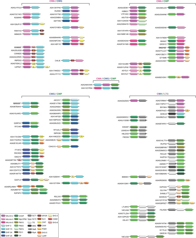
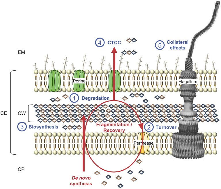
The cell wall (CW) of bacteria is an intricate arrangement of macromolecules, at least constituted of peptidoglycan (PG) but also of (lipo)teichoic acids, various polysaccharides, polyglutamate and/or proteins. During bacterial growth and division, there is a constant balance between CW degradation and biosynthesis. The CW is remodeled by bacterial hydrolases, whose activities are carefully regulated to maintain cell integrity or lead to bacterial death. Each cell wall hydrolase (CWH) has a specific role regarding the PG: (i) cell wall amidase (CWA) cleaves the amide bond between N-acetylmuramic acid and L-alanine residue at the N-terminal of the stem peptide, (ii) cell wall glycosidase (CWG) catalyses the hydrolysis of the glycosidic linkages, whereas (iii) cell wall peptidase (CWP) cleaves amide bonds between amino acids within the PG chain. After an exhaustive overview of all known conserved catalytic domains responsible for CWA, CWG, and CWP activities, this review stresses that the CWHs frequently display a modular architecture combining multiple and/or different catalytic domains, including some lytic transglycosylases as well as CW binding domains. From there, direct physiological and collateral roles of CWHs in bacterial cells are further discussed.

Keywords: bacterial cell wall; bacterial division and growth; cell lysis; cell wall binding domains; cell wall remodeling; peptidoglycan (PG) hydrolases; protein modules.


Figures

Figure 1
Figure 1
Schematic representation of the diversity of arrangements for bacterial cell envelopes. (A) Parietal monoderm (CW-monoderm) contains one cell membrane with a thick peptidoglycan layer and teichoic and lipoteichoic acids. (B) Lipopolysaccharidic diderm (LPS-diderm) bacteria are enveloped by two membranes with a thin peptidoglycan layer between the inner and outer membranes. (C) Mycolate diderm (myco-diderm) bacteria have a cytoplasmic membrane and a peculiar outer membrane constituted of mycolic acid, also called mycomembrane, with a cell wall in-between constituted of a peptidoglycan fraction and an arabinogalactan fraction. CE, cell envelope; CW, cell wall; CM, cytoplasmic membrane; OM, outer membrane; PS, periplasmic space; IM, inner membrane; IWZ, inner wall zone; MOM, mycolic outer membrane; PL, Phospholipid; PG, peptidoglycan; TA, teichoic acid; LTA, lipoteichoic acid; IMP, integral/inner membrane protein; IML, inner membrane lipoprotein; CWP, cell wall protein; OML, outer membrane lipoprotein; OMP, outer membrane protein; LPS, lipopolysaccharide; AG, arabinogalactan; GL, glycolipid; MA, mycolic acid; MOMP, mycolic outer membrane protein.
Figure 2
Figure 2
Targets of the CWHs in CW-monoderm and LPS-diderm bacteria. (A) In the CW of CW-monoderm bacteria, the alternating subunits of N-acetylglucosamine (GlcNac) and N-acetylmuramic acid (MurNac) are amide linked to the alanine of the wall peptide alanine, glutamine or isoglutamine, meso-diaminopimelic acid (mDAP) or lysine and alanine. In S. aureus, the pentaglycine cross bridge is linked to the alanine of the CW peptide. (B) In the CW of LPS-diderm bacteria, the alternating subunits of N-acetylglucosamine (GlcNac) and N-acetylmuramic acid (MurNac) are amide linked to the alanine of tetrapeptide alanine, glutamine, mDAP and alanine. N-acetylglucosaminidase hydrolyses the glycan component of the cell wall on the reducing side of the GlcNAc. In contrast, the lysozyme hydrolyses the glycan component of the cell wall on the reducing side of the MurNAc. Likewise, N-acetylmuramidases cleave the same bond but form N-acetyl-1,6-anhydro-muramyl intermediates during cleavage. N-acetylmuramoyl-L-alanine amidase cleaves a critical amide bond between the glycan moiety (MurNAc) and the peptide moiety (L-alanine) of the peptidoglycan. Peptidase cleaves an amide bond between two amino acids (depicted in green). CWA, CW amidase; CWG, CW glycosidase; CWP, CW peptidase.
Figure 3
Figure 3
Schematic representation of the diversity of modular architectures of CWHs acting as CWA only. For each modular organization, a UniProt identifier is provided as a representative. Bold identifier indicates that at least one representative protein has been functionally characterized (including the given identifier). Asterisk indicates that at least one representative has been structurally characterized (including the given identifier).
Figure 4
Figure 4
Schematic representation of the diversity of modular architectures of CWHs acting as CWG only. For each modular organization, a UniProt identifier is provided as a representative. Bold identifier indicates that at least one representative protein has been functionally characterized (including the given identifier). Asterisk indicates that at least one representative has been structurally characterized (including the given identifier).
Figure 5
Figure 5
Schematic representation of the diversity of modular architectures of CWHs acting as CWP only. For each modular organization, a UniProt identifier is provided as a representative. Bold identifier indicates that at least one representative protein has been functionally characterized (including the given identifier). Asterisk indicates that at least one representative has been structurally characterized (including the given identifier).
Figure 6
Figure 6
Schematic representation of the diversity of modular architectures of multifunctional CWHs, i.e., acting as CWA-CWG, CWA-CWP, CWG-CWP, CWA-CWG-CWP, or CWH-LTG. For each modular organization, a UniProt identifier is provided as a representative. Bold identifier indicates that at least one representative protein has been functionally characterized (including the given identifier). Asterisk indicates that at least one representative has been structurally characterized (including the given identifier).
Figure 7
Figure 7
Schematic representation of cell wall biosynthesis and recycling, exemplified in a LPS-diderm bacterial cell. Besides (1) degradation, which can lead to cell lysis, CWHs also participate to cell wall rearrangement and other key physiological functions, namely (2) turnover of cell wall material through their recycling to (3) biosynthesis, as well as (4) cell-to-cell communication (CTCC) since some released peptidoglycan fragments can act as signaling molecules or have (5) side effects on final protein subcellular localization, e.g., flagella, with consequences on motility, bacterial adhesion, biofilm formation, protein secretion, conjugation, virulence and/or immune response. CP, cytoplasm; CE, cell envelope; CW, cell wall; EM, extracellular milieu.

Similar articles

Cited by

References

    1. Abaev I., Foster-Frey J., Korobova O., Shishkova N., Kiseleva N., Kopylov P., et al. . (2013). Staphylococcal Phage 2638A endolysin is lytic for Staphylococcus aureus and harbors an inter-lytic-domain secondary translational start site. Appl. Microbiol. Biotechnol. 97, 3449–3456. 10.1007/s00253-012-4252-4 - DOI - PMC - PubMed
    1. Albrecht T., Raue S., Rosenstein R., Nieselt K., Götz F. (2012). Phylogeny of the staphylococcal major autolysin and its use in genus and species typing. J. Bacteriol. 194, 2630–2636. 10.1128/JB.06609-11 - DOI - PMC - PubMed
    1. Alcorlo M., Martinez-Caballero S., Molina R., Hermoso J. A. (2017). Carbohydrate recognition and lysis by bacterial peptidoglycan hydrolases. Curr. Opin. Struct. Biol. 44, 87–100. 10.1016/j.sbi.2017.01.001 - DOI - PubMed
    1. Alderwick L. J., Harrison J., Lloyd G. S., Birch H. L. (2015). The mycobacterial cell wall–peptidoglycan and arabinogalactan. Cold Spring Harb. Perspect. Med. 5:a021113. 10.1101/cshperspect.a021113 - DOI - PMC - PubMed
    1. Al-Riyami B., Ustok F. I., Stott K., Chirgadze D. Y., Christie G. (2016). The crystal structure of Clostridium perfringens SleM, a muramidase involved in cortical hydrolysis during spore germination. Proteins 84, 1681–1689. 10.1002/prot.25112 - DOI - PubMed

LinkOut - more resources