

Iron and Ferritin Modulate MHC Class I Expression and NK Cell Recognition
- PMID: 30873154
- PMCID: PMC6404638
- DOI: 10.3389/fimmu.2019.00224
Iron and Ferritin Modulate MHC Class I Expression and NK Cell Recognition
Abstract
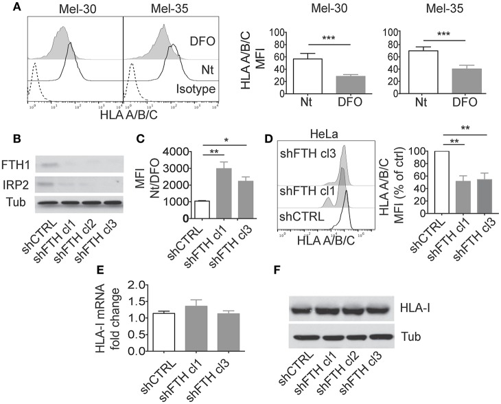
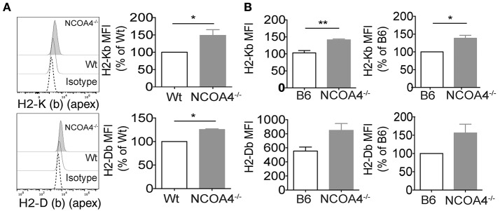
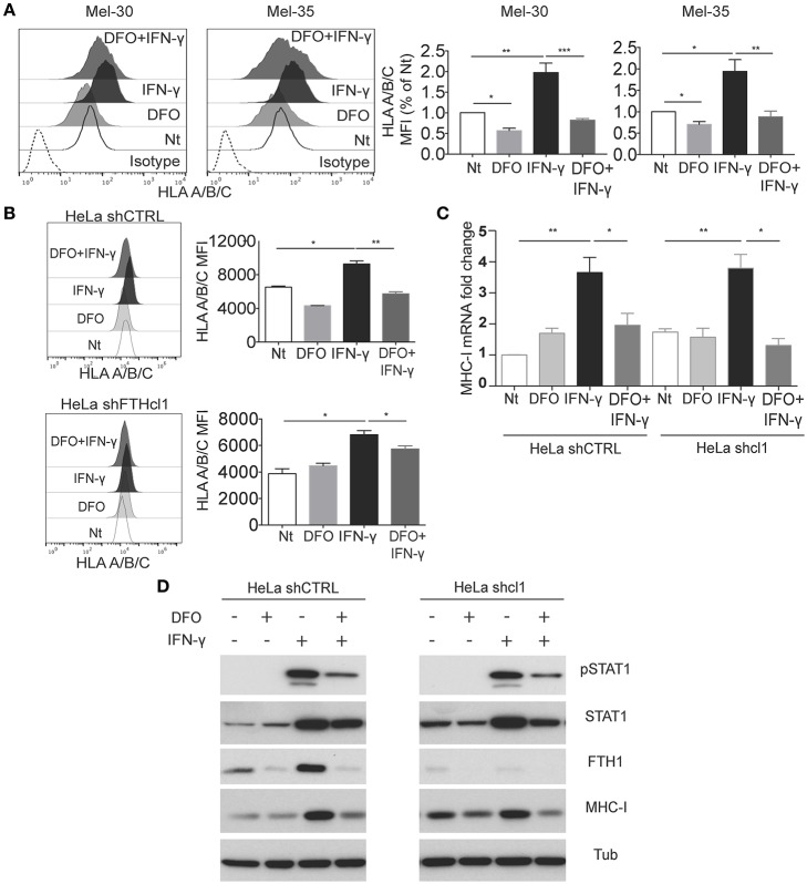
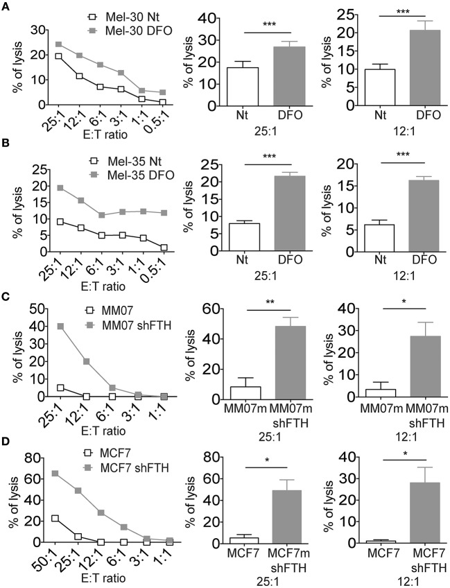
The ability of pathogens to sequester iron from their host cells and proteins affects their virulence. Moreover, iron is required for various innate host defense mechanisms as well as for acquired immune responses. Therefore, intracellular iron concentration may influence the interplay between pathogens and immune system. Here, we investigated whether changes in iron concentrations and intracellular ferritin heavy chain (FTH) abundance may modulate the expression of Major Histocompatibility Complex molecules (MHC), and susceptibility to Natural Killer (NK) cell cytotoxicity. FTH downregulation, either by shRNA transfection or iron chelation, led to MHC surface reduction in primary cancer cells and macrophages. On the contrary, mouse embryonic fibroblasts (MEFs) from NCOA4 null mice accumulated FTH for ferritinophagy impairment and displayed MHC class I cell surface overexpression. Low iron concentration, but not FTH, interfered with IFN-γ receptor signaling, preventing the increase of MHC-class I molecules on the membrane by obstructing STAT1 phosphorylation and nuclear translocation. Finally, iron depletion and FTH downregulation increased the target susceptibility of both primary cancer cells and macrophages to NK cell recognition. In conclusion, the reduction of iron and FTH may influence the expression of MHC class I molecules leading to NK cells activation.
Keywords: HLA; IFNγ; MHC-I; NK cells; STAT1; iron.
Figures
Similar articles
-
Protection against natural killer cells by interferon-gamma treatment of K562 cells cannot be explained by augmented major histocompatibility complex class I expression.Immunology. 1994 Sep;83(1):75-80. Immunology. 1994. PMID: 7821970 Free PMC article.
-
NK cells modulate MHC class I expression on tumor cells and their susceptibility to lysis.Immunobiology. 2000 Nov;202(4):326-38. doi: 10.1016/s0171-2985(00)80037-6. Immunobiology. 2000. PMID: 11131150
-
[Expression of HLA class I molecules and MHC class I chain-related molecules A/B in K562 and K562/AO2 cell lines and their effects on cytotoxicity of NK cells].Zhongguo Shi Yan Xue Ye Xue Za Zhi. 2007 Apr;15(2):288-91. Zhongguo Shi Yan Xue Ye Xue Za Zhi. 2007. PMID: 17493333 Chinese.
-
Histocompatibility antigens and natural killer susceptibility.Immunol Res. 1992;11(2):133-40. doi: 10.1007/BF02918618. Immunol Res. 1992. PMID: 1431422 Review.
-
Dual effects of cytokines in regulation of MHC-unrestricted cell mediated cytotoxicity.Crit Rev Immunol. 1993;13(1):1-34. Crit Rev Immunol. 1993. PMID: 8466640 Review.
Cited by
-
In the SARS-CoV-2 Pandora Pandemic: Can the Stance of Premorbid Intestinal Innate Immune System as Measured by Fecal Adnab-9 Binding of p87:Blood Ferritin, Yielding the FERAD Ratio, Predict COVID-19 Susceptibility and Survival in a Prospective Population Database?Int J Mol Sci. 2023 Apr 19;24(8):7536. doi: 10.3390/ijms24087536. Int J Mol Sci. 2023. PMID: 37108697 Free PMC article.
-
The Iron Curtain: Macrophages at the Interface of Systemic and Microenvironmental Iron Metabolism and Immune Response in Cancer.Front Immunol. 2021 Apr 27;12:614294. doi: 10.3389/fimmu.2021.614294. eCollection 2021. Front Immunol. 2021. PMID: 33986740 Free PMC article. Review.
-
Emerging role of ferroptosis in glioblastoma: Therapeutic opportunities and challenges.Front Mol Biosci. 2022 Aug 17;9:974156. doi: 10.3389/fmolb.2022.974156. eCollection 2022. Front Mol Biosci. 2022. PMID: 36060242 Free PMC article. Review.
-
Therapeutic potential of induced iron depletion using iron chelators in Covid-19.Saudi J Biol Sci. 2022 Apr;29(4):1947-1956. doi: 10.1016/j.sjbs.2021.11.061. Epub 2021 Dec 13. Saudi J Biol Sci. 2022. PMID: 34924800 Free PMC article. Review.
-
FtH-Mediated ROS Dysregulation Promotes CXCL12/CXCR4 Axis Activation and EMT-Like Trans-Differentiation in Erythroleukemia K562 Cells.Front Oncol. 2020 May 5;10:698. doi: 10.3389/fonc.2020.00698. eCollection 2020. Front Oncol. 2020. PMID: 32432042 Free PMC article.
References
Publication types
MeSH terms
Substances
LinkOut - more resources
Full Text Sources
Other Literature Sources
Medical
Research Materials
Miscellaneous