. 2019 Feb 27:10:330.
doi: 10.3389/fimmu.2019.00330. eCollection 2019.

Persistence of Candida albicans in the Oral Mucosa Induces a Curbed Inflammatory Host Response That Is Independent of Immunosuppression

Affiliations

Persistence of Candida albicans in the Oral Mucosa Induces a Curbed Inflammatory Host Response That Is Independent of Immunosuppression

Florian R Kirchner et al. Front Immunol. .

Abstract

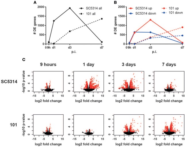
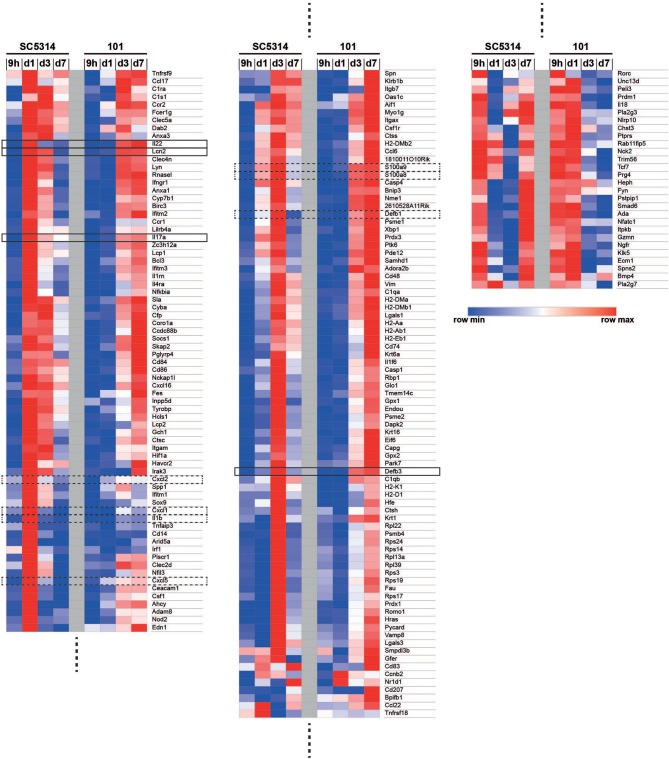
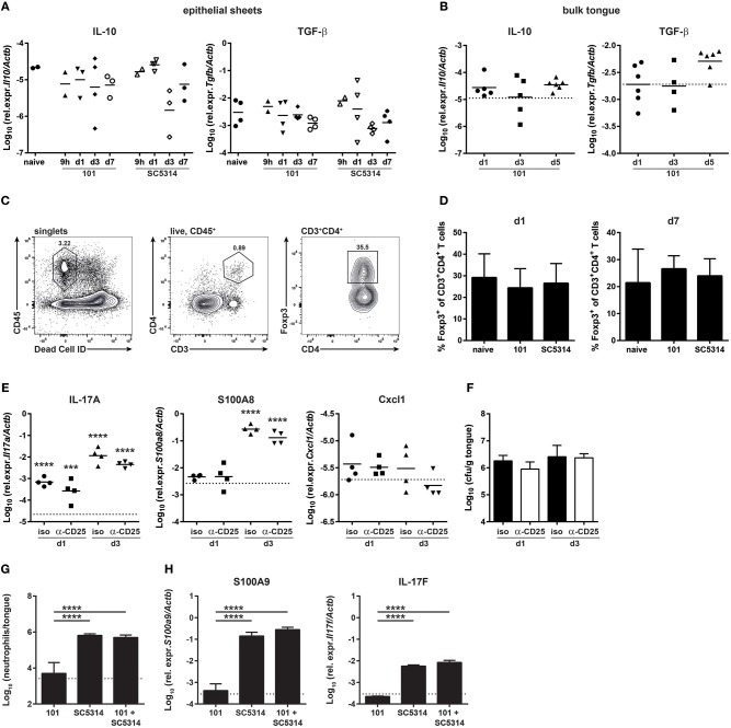
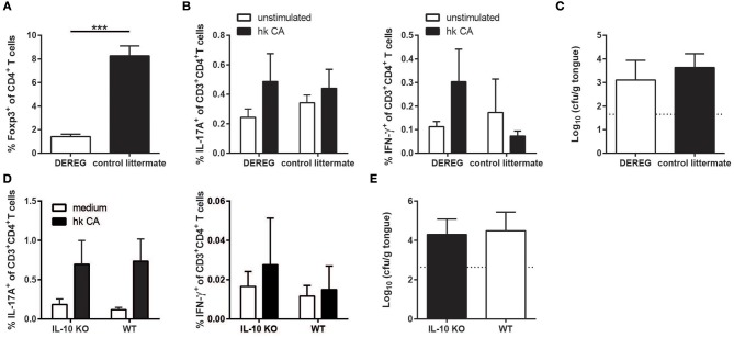
Controlled immune activation in response to commensal microbes is critical for the maintenance of stable colonization and prevention of microbial overgrowth on epithelial surfaces. Our understanding of the host mechanisms that regulate bacterial commensalism has increased substantially, however, much less data exist regarding host responses to members of the fungal microbiota on colonized surfaces. Using a murine model of oropharyngeal candidiasis, we have recently shown that differences in immune activation in response to diverse natural isolates of Candida albicans are associated with different outcomes of the host-fungal interaction. Here we applied a genome-wide transcriptomic approach to show that rapid induction of a strong inflammatory response characterized by neutrophil-associated genes upon C. albicans colonization inversely correlated with the ability of the fungus to persist in the oral mucosa. Surprisingly, persistent fungal isolates showed no signs of a compensatory regulatory immune response. By combining RNA-seq data, genetic mouse models, and co-infection experiments, we show that attenuation of the inflammatory response at the onset of infection with a persistent isolate is not a consequence of enhanced immunosuppression. Importantly, depletion of regulatory T cells or deletion of the immunoregulatory cytokine IL-10 did not alter host-protective type 17 immunity nor did it impair fungal survival in the oral mucosa, indicating that persistence of C. albicans in the oral mucosa is not a consequence of suppressed antifungal immunity.

Keywords: Candida albicans; IL-10; IL-17; immune regulation; oropharyngeal candidiasis; persistence; regulatory T cells.


Figures

Figure 1
Figure 1
The host response to C. albicans strain 101 is delayed compared to strain SC5314. Epithelial sheet from C. albicans strain SC5314- and strain 101-infected WT mice were subjected to RNA-seq analysis. (A) Graph showing the total number of differentially expressed (DE) genes at the indicated time points compared to naïve controls. (B) Separate display of the up- and down-regulated genes at the indicated time points. (C) Volcano plots displaying the fold changes and the FDR of all genes detected in each condition separately. Genes with FDR < 0.05 and fold change < −2 or > 2 are marked in red.
Figure 2
Figure 2
Co-regulated and differentially regulated genes in the mucosal response to strains 101 and SC5314. The GO term enrichment analysis on genes that are differentially regulated in at least one contrast for the 62 GO terms that are offspring of GO: 0002376 (immune system processes) with p < 0.05 yielded 182 genes. Genes were ordered by hierarchical clustering of their log2 fold changes, which are displayed in a heat map with a row-scaling color scheme.
Figure 3
Figure 3
The persistent strain 101 does not suppress the antifungal host response. (A,B) Relative expression of Il10 (left) and Tgfb1 transcripts (right) in epithelial sheets (A) or in bulk tongue tissue (B) of mice that were infected with strain 101 or SC5314 for the indicated period of time. Each symbol represents a pool of epithelial sheets from three animals each (A) or a single mouse (B). The geomean of each group is indicated. Data are pooled from two independent experiments each. (C,D) % Foxp3+ Tregs within the viable CD45+CD3+CD4+ population in the tongue of mice that were infected with strain 101 or SC5314 for the indicated period of time. Representative FACS plots and the gating strategy are shown in C, summary plots showing the mean + SD of data pooled from two independent experiments with 3–4 animals per group are shown in D. (E,F) Mice were treated with anti-CD25 or isotype control antibody prior to infection with strain 101. Relative expression of Il17a (left), S100a8 (middle), and Cxcl1 (right) (E) and tongue fungal loads (F) in the bulk tongue tissue at the indicated time point after infection are shown. In (E), each symbol represents a single animal, The geomean of each group is indicated. The dotted line represents transcript levels in naïve animals (mean of 8 animals). In (F), each bar is the geomean + SD of 4 animals per group. (G,H) WT mice were infected sublingually with a 1:1 mixture of strain 101 and strain SC5314 or with each strain alone. Tongues were harvested on day 1 post-infection and analyzed for the infiltration of Ly6G+Ly6CloCD11b+ neutrophils by flow cytometry (G) and for the expression of S100a9 and Il17f (H) transcript by RT-qPCR. Data are the mean + SD of 3–4 mice per group. Graphs display data representative of one out of two independent experiments. The dotted line represents the detection limit. Statistics were calculated using one-way ANOVA. In (E), statistics compare infected to naïve groups. ***p < 0.001, ****p < 0.0001.
Figure 4
Figure 4
The Treg response during persistent colonization of the oral mucosa with C. albicans. (A–F) WT mice were sublingually infected with strain 101 or SC5314 and cervical lymph node cells were analyzed on day 7 or day 18–21 as indicated. (A,B) Lymph node cells were re-stimulated with MutuDC1940 cells that were pulsed with heat-killed C. albicans or left unpulsed for 5 h in the presence of Brefeldin A. IL-17A production by CD3+CD4+ cells was analyzed by intracellular cytokine staining and flow cytometry. (C,D) The frequency of Foxp3-expressing cells within the CD4+ lymphocyte compartment was assessed by flow cytometry. (E,F) PD-1, TIGIT, and Tim-3 expression by CD4+Foxp3+ Treg cells was analyzed by flow cytometry. (G–J) Il10-Thy1.1 reporter mice were sublingually infected with strain 101 or SC5314 or left naive. IL-10 expression by Foxp3+ Tregs and Foxp3 effector T cells was assessed in the cervical lymph nodes (G,H) and in the tongue (I,J) on day 21 post-infection by flow cytometry. Cells were pregated on CD90+CD4+ (G,H) or on CD45.2+CD3+ (I,J), respectively. Representative FACS plots are shown in (A, C, G, I); summary plots with data pooled from at least 2 experiments with 6–9 animals per infected group and 3–4 animals per naive group are shown in (B, D–F, H, J), with the exception of the right plot in (D), where data are from a single experiment with 3 animals per group. In B, Statistics were calculated using t-test. In (D–F, H, J), statistics were calculated using one-way ANOVA. *p < 0.05.
Figure 5
Figure 5
The absence of Tregs or IL-10 does not compromise persistence of strain 101 in the oral epithelium. (A–C) DEREG mice and control littermates were sublingually infected with C. albicans strain 101 and treated with diphtheria toxin on day 11 and 13 after the antifungal response was fully established. One day after the last treatment, the mice were sacrificed for analysis. (A) Treg depletion efficiency was analyzed in the cervical lymph nodes by flow cytometry. Data are the % of Foxp3+ cells within the population of CD4+ viable cells. (B) Lymph node cells were re-stimulated with MutuDC1940 cells that were pulsed with heat-killed C. albicans or left unpulsed for 5 h in the presence of Brefeldin A. IL-17A (left) and IFN-γ (right) production by CD3+CD4+ cells was analyzed by intracellular cytokine staining and flow cytometry. (C) The fungal burden was determined by plating tongue homogenates on YPD agar. Each bar represents the mean + SD of 3 to 4 mice per group. Data are from one out of two independent experiments. (D,E) IL-10-deficient mice and WT controls were sublingually infected with C. albicans strain 101 and analyzed on day 9 post-infection. (D) Lymph node cells were re-stimulated and analyzed for IL-17 (left) and IFN-γ (right) production as in B. (E) Tongue fungal burdens were analyzed as in C. Each bar represents the mean + SD of 8–9 mice per group pooled from two independent experiments. Statistics were calculated using unpaired t-Test. ***p < 0.001.

References

    1. Brown GD, Denning DW, Gow NA, Levitz SM, Netea MG, White TC. Hidden killers: human fungal infections. Sci Transl Med. (2012) 4:165rv13. 10.1126/scitranslmed.3004404 - DOI - PubMed
    1. de Repentigny, Lewandowski D, Jolicoeur P. Immunopathogenesis of oropharyngeal candidiasis in human immunodeficiency virus infection. Clin Microbiol Rev. (2004) 17:729–59, table of contents. 10.1128/CMR.17.4.729-759.2004 - DOI - PMC - PubMed
    1. Goncalves B, Ferreira C, Alves CT, Henriques M, Azeredo J, Silva S. Vulvovaginal candidiasis: epidemiology, microbiology and risk factors. Crit Rev Microbiol. (2016) 42:905–27. 10.3109/1040841X.2015.1091805 - DOI - PubMed
    1. Kirkpatrick CH. Chronic mucocutaneous candidiasis. Pediatr Infect Dis J. (2001) 20:197–206. 10.1097/00006454-200102000-00017 - DOI - PubMed
    1. Puel A, Cypowyj S, Marodi L, Abel L, Picard C, Casanova JL. Inborn errors of human IL-17 immunity underlie chronic mucocutaneous candidiasis. Curr Opin Allergy Clin Immunol. (2012) 12:616–22. 10.1097/ACI.0b013e328358cc0b - DOI - PMC - PubMed

Publication types

MeSH terms