. 2019 Feb 25:7:72.
doi: 10.3389/fchem.2019.00072. eCollection 2019.

Dynamic Control of the Self-Assembling Properties of Cyclodextrins by the Interplay of Aromatic and Host-Guest Interactions

Affiliations

Dynamic Control of the Self-Assembling Properties of Cyclodextrins by the Interplay of Aromatic and Host-Guest Interactions

Tania Neva et al. Front Chem. .

Abstract

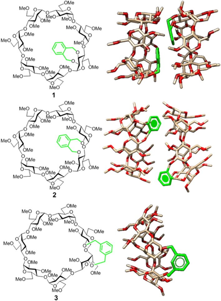
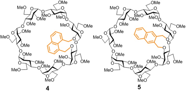
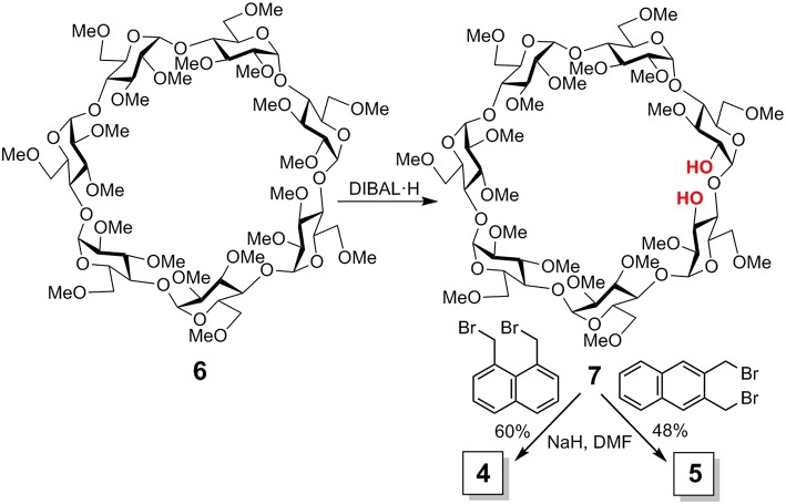
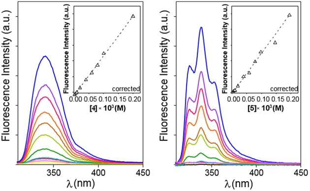
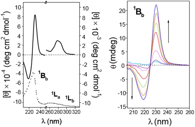
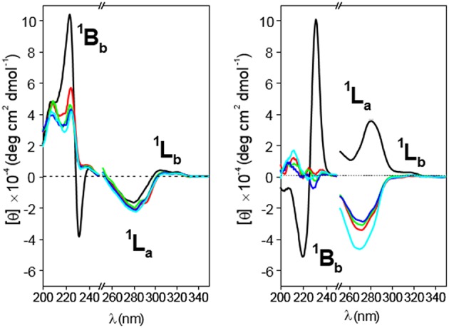
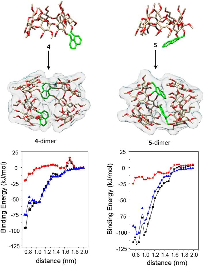
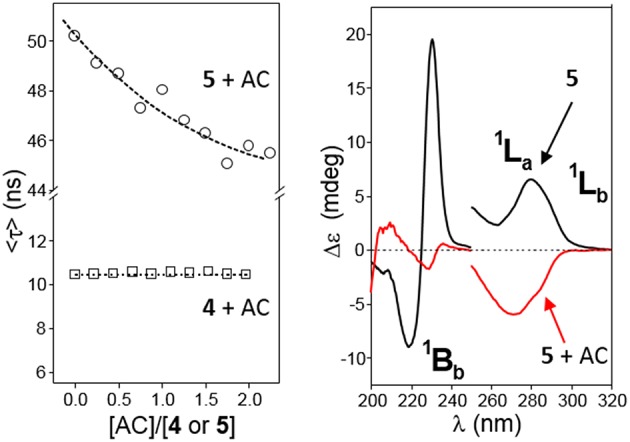
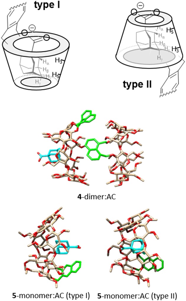
The presence of a doubly-linked naphthylene clip at the O-2I and O-3II positions in the secondary ring of β-cyclodextrin (βCD) derivatives promoted their self-assembly into head-to-head supramolecular dimers in which the aromatic modules act either as cavity extension walls (if the naphthalene moiety is 1,8-disubstituted) or as folding screens that separate the individual βCD units (if 2,3-disubstituted). Dimer architecture is governed by the conformational properties of the monomer constituents, as determined by NMR, fluorescence, circular dichroism, and computational techniques. In a second supramolecular organization level, the topology of the assembly directs host-guest interactions and, reciprocally, guest inclusion impacts the stability of the supramolecular edifice. Thus, inclusion of adamantane carboxylate, a well-known βCD cavity-fitting guest, was found to either preserve the dimeric arrangement, leading to multicomponent species, or elicit dimer disruption. The ensemble of results highlights the potential of the approach to program self-organization and external stimuli responsiveness of CD devices in a controlled manner while keeping full diastereomeric purity.

Keywords: circular dichroism; cyclodextrins; fluorescence; host-guest chemistry; naphthalene; self-assembly; supramolecular chemistry.


Figures

Figure 1
Figure 1
Structures of the doubly-linked isomeric βCD-xylylene conjugates 13 (Left) and 3D molecular models of the corresponding minima binding energy dimer (for 1 and 2) or monomer species (for 3) present in water solution (Right). Note that the aromatic ring (in green) orientation, relative to the CD cavity, shifts from cap-like to semi-open to fully open on moving from 1 to 3 (González-Álvarez et al., ; Neva et al., 2018).
Figure 2
Figure 2
Structures of the new βCD conjugates equipped with naphthalene clips prepared in this work.
Figure 3
Figure 3
Synthesis of compounds 4 and 5.
Figure 4
Figure 4
Emission spectra for 4 (Left) and 5 (Right) in water in the 0 to ~0.2 mM range of concentrations (yellow to blue solid lines) at 25°C. Variation of the corrected fluorescence intensity (Equation S1) with compound concentration measured at the intensity maxima are superimposed (dashed lines).
Figure 5
Figure 5
Variation of the weighted average lifetime <τ> with the concentration of 4 (Left) or 5 (Right) in water in the 5–45°C temperature range.
Figure 6
Figure 6
Van't Hoff linear representations for the dimerization processes of 4 and 5 obtained from the association constants collected in Table 1.
Figure 7
Figure 7
Absorption spectra for 4 (Top Left) and 5 (Top Right) in water at 25°C, with the corresponding absorbance vs. concentration plots (inserts) and directions of the electronic transition moments for the 1,8- (Bottom Left) and 2,3-naphthylene (Bottom Right) appended groups.
Figure 8
Figure 8
(Left) 210–330 nm range circular dichroism spectrum (solid line; as molar ellipticity [θ]), and absorption spectra (dotted line; arbitrary units) for 4 (0.11 mM in water). (Middle and Right) Circular dichroism spectra (as ellipticity; θ) for 4 in the 200–260 nm (from magenta to blue lines: 0.67, 1.12, 1.68 2.24, and 3.36 · 10−4 M in water; cell path 0.1 cm) and in the 260–330 nm range (from yellow to blue lines: 0.022 · 10−4 to 3.4 · 10−4 M in water; cell path 1 cm). All spectra were recorded at 25°C.
Figure 9
Figure 9
Circular dichroism spectra of aqueous solutions of CD 5. (Left) Represents molar ellipticity ([θ], solid lines) and pure absorption (dotted lines) of a 0.30 mM solution in the 210–330 nm range. (Right) Represents the ellipticity (θ) at variable concentration (from light blue to blue lines: 0.17, 0.26, 0.54, 1.09, 1.63, and 2.73 · 10−4 M) in the 200–260 nm (path 0.1 cm). All spectra recorded at 25°C.
Figure 10
Figure 10
Circular dichroism spectra for 4 (Left) and 5 (Right) solutions in water (black line), 1:1 (v/v) water/methanol (red line), methanol (green line), ethanol (deep blue line), and propanol (light blue line) at 25°C. Concentrations were fixed at 0.19 and 0.20 mM for 4 and 5, respectively.
Figure 11
Figure 11
The (Top and Middle) represent 3D-views of the optimized (MM) structures of 4 and 5 (oxygen in red, carbon in tan; H atoms have been removed and the aromatic ring is highlighted in green for the sake of clarity) and minima binding energy (MBE) structures of the corresponding HH-dimers. The (Bottom) contains the changes of the total interaction energies (black) as well as the electrostatics (red) and van der Waals (blue) contributions, obtained after step by step approaching along the y axis defined by the center of mass of the glycosidic oxygens (see the Experimental section and the Supplementary Materials for details).
Figure 12
Figure 12
(Left) Plots of the average lifetime (<τ>) variations for water solutions of 4 (□) or 5 (◦, 1.36 · 10−4 M) at increasing AC concentrations at 25°C. (Right) Circular dichroism spectra of 5 (0.15 mM in water) in the absence (black line) and in the presence (red line) of a large excess of AC ([AC]/[5] = 86) at 25°C.
Figure 13
Figure 13
(Top) Schematic representation of type I and type II complexes between AC and βCD derivatives bearing aromatic clips, with indication of the expected diagnostic NOE contacts. (Middle) MBE structure of the ternary complex formed by 4-dimer and AC. (Bottom) MBE structures of the type I (left) and type II (right) 5-dimer:AC complexes obtained by approaching the AC guest to the CD host by the primary rim, to give a type I complex (left), or by the secondary rim, to give a type II complex (right). In the CD partners, oxygens are colored in red, carbons in tan; H atoms have been removed and the aromatic ring is highlighted in green; in the AC partner, the carbon atoms are highlighted in cyan for the sake of clarity.

Similar articles

Cited by

References

    1. Álvarez-Dorta D., León E. I., Kennedy A. R., Martín A., Pérez-Martín A., Suárez E. (2016). Radical-mediated C–H functionalization: a strategy for access to modified cyclodextrins. J. Org. Chem. 81, 11766–11787. 10.1021/acs.joc.6b02241 - DOI - PubMed
    1. Álvarez-Dorta D., León E. I., Kennedy A. R., Martín A., Pérez-Martín I., Suárez E. (2015). Easy access to modified cyclodextrins by an intramolecular radical approach. Angew. Chem. Int. Ed. 54, 3674–3678. 10.1002/anie/201412300 - DOI - PubMed
    1. Álvarez-Lorenzo C., García-González C. A., Concheiro A. (2017). Cyclodextrins as versatile building blocks for regenerative medicine. J. Control. Release 268, 269–281. 10.1016/j.jconrel.2017.10.038 - DOI - PubMed
    1. Aoyagi T., Nakamura A., Ikeda H., Ikeda T., Mihara H., Ueno A. (1997). Alizarin Yellow-modified? β-cyclodextrin as a guest-responsive absorption change sensor. Anal. Chem. 69, 659–663. 10.1021/ac960727z - DOI
    1. Aranda C., Urbiola K., Méndez Ardoy A., García Fernández J. M., Ortiz Mellet C., Tros de Ilarduya C. (2013). Targeted gene delivery by new folate–polycationic amphiphilic cyclodextrin–DNA nanocomplexes in vitro and in vivo. Eur. J. Pharm. Bipharm. 85, 390–397. 10.1016/j.ejpb.2013.06.011 - DOI - PubMed

LinkOut - more resources