Review
. 2015 Sep 30;1(2):94-99.

Diagnosis and treatment of uveitis; not restricted to the ophthalmologist

Affiliations
Review

Diagnosis and treatment of uveitis; not restricted to the ophthalmologist

Jan A M van Laar et al. J Clin Transl Res. .

Abstract

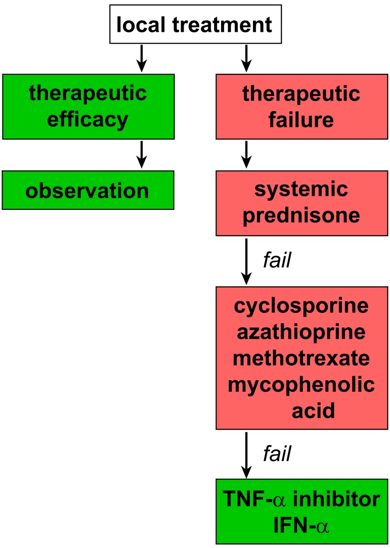
Uveitis is associated with a wide range of underlying causes. Familiarity with its clinical manifestations, referral indications, and treatment strategies are required for the optimal use of current therapeutic options. Uveitis can be caused by infectious and non-infectious factors, resulting in differing prognoses and treatments. The treatment of chronic, non-infectious uveitis has profoundly changed in the last years due to the advent of biologicals, but also of intraocular therapies. In severe uveitis, treatment of the underlying cause, whether ocular or systemic, is required to prevent severe loss of vision. For these purposes, a multidisciplinary clinical approach is important, which is addressed in this review. Relevance for patients: A broad understanding of the different causes of uveitis and the implementation of disease-tailored, multidisciplinary management of uveitis is expected to improve treatment outcomes for patients with different types of uveitis.

Keywords: anti-TNF; ethiology; immunosuppressive therapy; multidisciplinary management; uveitis.


Conflict of interest statement

The authors declare that there are no conflicts of interest present.
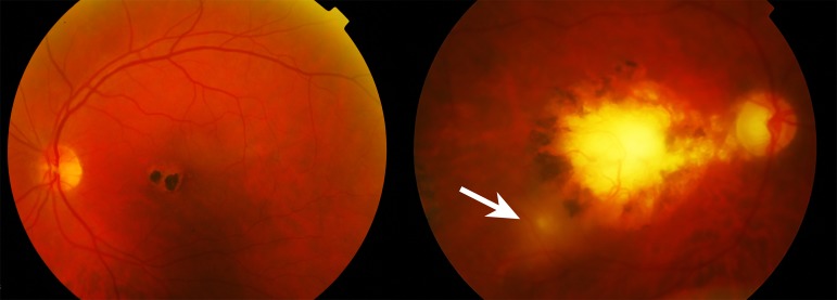
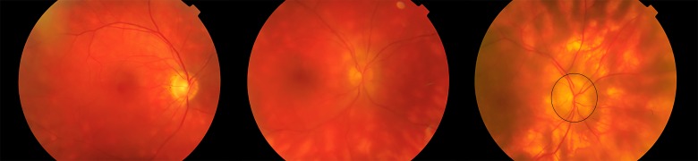
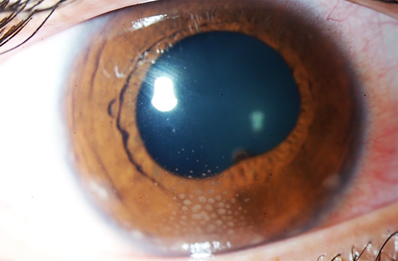
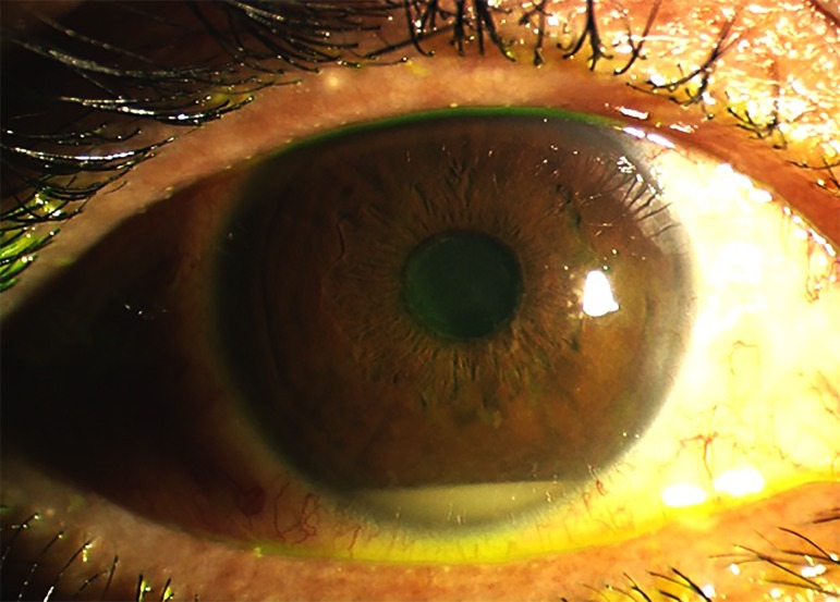
Figures

Figure 1.
Figure 1.. Color fundus photograph of chorioretinitis due to a toxoplasmosis infection. The left panel presents the left eye showing a central scar in the macula with both hypo- and hyperpigmentation. The right panel depicts exacerbation of active inflammation (hazy yellowish area at the edge of the pigmented scar (arrow)). The haziness of the right panel is attributed to inflammation of the vitreous.
Figure 2.
Figure 2.. Color fundus photographs of the right eye of different patients with ‘Birdshot’ chorioretinitis, showing the characteristic multiple, yellow chorioretinal inflammatory lesions with increasing severity (from left to right). The right panel also shows mild swelling of the optic disc (encircled).
Figure 3.
Figure 3.. Color image of the anterior segment of the right eye of a patient with granulomatous anterior uveitis. Typical for this presentation are the mutton-fat keratic precipitates organized in ‘Arlt’s triangle’ (approximately 6 o’clock) on the interior surface of the cornea (endothelium). The image additionally shows mild ciliary hyperemia of the conjunctiva and adhesion of the iris to the lens capsule (pupil margin at approximately 5 o’clock).
Figure 4.
Figure 4.. Color of the anterior segment of a patient with Behçet’s disease. Accumulation of granulocytes creating a liquid level in the anterior chamber is clearly visible. Although hypopyon is an archetypal ocular feature of Behçet’s disease, the visual prognosis is determined by changes in the posterior eye segment, including injury to the macula and optic nerve.
Figure 5.
Figure 5.. Flow diagram for the management of non-infectious uveitis.

Similar articles

Cited by

References

    1. Jabs DA, Nussenblatt RB, Rosenbaum JT. Standardization of uveitis nomenclature for reporting clinical data. Results of the First International Workshop. Am J Ophthalmol. 2005;140:509–516. - PMC - PubMed
    1. Gritz DC, Wong IG. Incidence and prevalence of uveitis in Northern California; the Northern California Epidemiology of Uveitis Study. Ophthalmology. 2004;111:491–500. - PubMed
    1. Lardenoye CW, van KB, Rothova A. Impact of macular edema on visual acuity in uveitis. Ophthalmology. 2006;113:1446–1449. - PubMed
    1. Durrani OM, Tehrani NN, Marr JE, Moradi P, Stavrou P, Murray PI. Degree, duration, and causes of visual loss in uveitis. Br J Ophthalmol. 2004;88:1159–1162. - PMC - PubMed
    1. Westeneng AC, Rothova A, de Boer JH, de Groot-Mijnes JD. Infectious uveitis in immunocompromised patients and the diagnostic value of polymerase chain reaction and Goldmann-Witmer coefficient in aqueous analysis. Am J Ophthalmol. 2007;144:781–785. - PubMed

LinkOut - more resources