Review
. 2019 Mar;7(2):10.1128/microbiolspec.gpp3-0039-2018.
doi: 10.1128/microbiolspec.GPP3-0039-2018.

Staphylococcus aureus Secreted Toxins and Extracellular Enzymes

Affiliations
Review

Staphylococcus aureus Secreted Toxins and Extracellular Enzymes

Kayan Tam et al. Microbiol Spectr. 2019 Mar.

Abstract

Staphylococcus aureus is a formidable pathogen capable of causing infections in different sites of the body in a variety of vertebrate animals, including humans and livestock. A major contribution to the success of S. aureus as a pathogen is the plethora of virulence factors that manipulate the host's innate and adaptive immune responses. Many of these immune modulating virulence factors are secreted toxins, cofactors for activating host zymogens, and exoenzymes. Secreted toxins such as pore-forming toxins and superantigens are highly inflammatory and can cause leukocyte cell death by cytolysis and clonal deletion, respectively. Coagulases and staphylokinases are cofactors that hijack the host's coagulation system. Exoenzymes, including nucleases and proteases, cleave and inactivate various immune defense and surveillance molecules, such as complement factors, antimicrobial peptides, and surface receptors that are important for leukocyte chemotaxis. Additionally, some of these secreted toxins and exoenzymes can cause disruption of endothelial and epithelial barriers through cell lysis and cleavage of junction proteins. A unique feature when examining the repertoire of S. aureus secreted virulence factors is the apparent functional redundancy exhibited by the majority of the toxins and exoenzymes. However, closer examination of each virulence factor revealed that each has unique properties that have important functional consequences. This chapter provides a brief overview of our current understanding of the major secreted virulence factors critical for S. aureus pathogenesis.


Figures

FIGURE 1
FIGURE 1
Current models for PFT pore formation for (A) α-toxin and (B) the bicomponent PFTs. (A) α-toxin is secreted as a monomer. Upon binding to the host receptor, ADAM-10, the toxin monomers oligomerize to form a heptameric prepore on the target cell surface. The prestem domains of the prepore then extend to form a β-barrel pore that punctures the target cell membrane. (B) The bicomponent PFTs are also secreted as monomers (except LukAB, which is secreted as dimers). The S-subunit recognizes the target cell by binding to cell surface receptors (LukPQ is an exception; the F-subunit LukQ is the receptor recognition subunit). These receptors are typically G-protein-coupled receptors (except for LukAB, which binds to the integrin CD11b). Upon receptor binding, the S-subunit dimerizes with the F-subunit, followed by oligomerization of three additional leukocidin dimers, resulting in an octameric prepore. Similar to the α-toxin pore formation model, the prestem domains of the prepore extend to form a β-barrel pore, thus disrupting the target cell membrane.
FIGURE 2
FIGURE 2
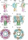
Structures of (A to C) α-toxin and (D to G) the bicomponent PFT, HlgAB. (A) The α-toxin monomer (PDB:4U6V) (351). The amino latch is colored blue, the cap domain red, the rim domain pink, and the prestem domain green. (B, C) The α-toxin heptamer (7AHL) (4). Each α-toxin is colored a different shade of pink to denote individual protomers. The amino latches are highlighted in blue, and the β-barrel pore is green. The monomers of (D) HlgA (2QK7) (352) and (E) HlgB (1LKF) (353). The amino latch of HlgB is blue; the cap domain for HlgA is cyan and HlgB is beige; the rim domains are yellow for HlgA and pink for HlgB; and the prestem domains are green. (F, G) The HlgAB octamer (3B07) (33). The HlgA protomers are cyan, the HlgB protomers are beige, and the β-barrel pore is green.
FIGURE 3
FIGURE 3
S. aureus PFTs and their receptor, species, and cell type specificities. Currently, S. aureus is known to produce eight β-barrel PFTs. Each of these PFTs targets different cell surface receptors. While some PFTs share the same receptors, they can differ in their species specificity. Collectively, the PFTs exert their sublytic and lytic effects on a variety of cells, including erythrocytes, endothelial cells, epithelial cells, neutrophils, monocytes, macrophages, dendritic cells, and T cells.
FIGURE 4
FIGURE 4
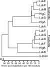
Phylogenic tree of S. aureus PFTs. The tree is constructed based on the mature protein sequences using the DNASTAR MegAlign ClustalW method for multiple sequence alignment.
FIGURE 5
FIGURE 5
Phylogenic tree of S. aureus SAgs. The tree is constructed based on the mature protein sequences using the DNASTAR MegAlign ClustalW method for multiple sequence alignment.
FIGURE 6
FIGURE 6
Crystal structures of S. aureus superantigens in complex with their cellular targets. (A) The T cell superantigen, SEB, in complex with the TCR and MHC II molecule (4C56) (116). SEB (blue) cross-links the α-chain of MHC (dark green) to the Vβ TCR (orange) to induce T cell proliferation that results in T cell anergy and/or apoptosis. (B) The B cell superantigen, SpA (teal), in complex with the Fab fragment (pink/magenta) (1DEE) (138). Conventional antigens bind to B cell receptors at the complementarity-determining region (blue), a hypervariable region that confers antigen specificities. SpA binds at a constant region of the receptor to activate B cells for supraclonal expansion, which leads to clonal deletion of SpA-activated B cells.
FIGURE 7
FIGURE 7
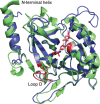
Overlay of the crystal structures of ETA and ETB. ETA (1EXF, green) (174) and ETB (1QTF, blue) (173) share high structural identity. ETs cause SSSS by cleaving Dsg1 at the epithelial cell junctions. Both ETs are serine proteases. Loop D and the catalytic triad are highlighted in pink for ETA and red for ETB.
FIGURE 8
FIGURE 8
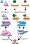
S. aureus produces cofactors that hijack the host’s coagulation system. Coa and vWbp bind to prothrombin and alter the conformation of the protein to form the complex, staphylothrombin. This complex is highly active and cleaves fibrinogens to fibrins, promoting the formation of fibrinous clots. Sak binds to plasmin to form the Sak-plasmin complex. Sak stabilizes plasmin to enhance enzymatic activity. Sak-plasmin cleaves plasminogen to form plasmin, which breaks down fibrin clots.
FIGURE 9
FIGURE 9
The crystal structure of Sak in complex with two plasminogen molecules (1BUI) (215). While Sak binding to plasminogen does not have enzymatic activity, the trimeric complex captures how Sak may bind to plasmin to cleave plasminogen. Sak (orange) is in complex with plasminogen (blue), exposing the catalytic site (red). Sak facilitates the docking of the substrate plasminogen (pink) to promote cleavage by plasmin.
FIGURE 10
FIGURE 10
Staphylococcal protease cascade. The metalloprotease, Aur, is activated by autoproteolysis after protein secretion. Aur is required to activate the serine protease, SspA. SspA processes one of the staphopains, SspB, from zymogen to active enzyme. The other staphopain, ScpA, is activated by autoproteolysis. Both staphopains are inhibited by staphostatins prior to secretion. SspB is inhibited by SspC, and ScpA is inhibited by ScpB.
FIGURE 11
FIGURE 11
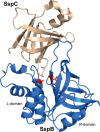
Staphopain-staphostatin complex (1PXV) (293). Staphopain SspB (blue) has two domains: the L-domain is helical, and the R-domain consists of β-strands that fold into a β-barrel-like structure. The catalytic site of SspB is highlighted in red. Staphopain SspC (beige) is a single domain protein composed of eight β-strands forming a single mixed β-barrel domain. SspC is a competitive inhibitor of SspB, directly blocking substrate access to the active site.
FIGURE 12
FIGURE 12
(A) Phylogenic tree of S. aureus Spls. The tree is constructed based on the mature protein sequences using the DNASTAR MegAlign ClustalW method for multiple sequence alignment. (B) Crystal structure of SplA (2W7S) (299). SplA has two domains connected by a linker (cyan). Domain 1 (light purple) consists of α-helices and β-strands, and domain 2 (blue) consists of of β-strands. Both domains fold into a β-barrel structure. The catalytic triad (red) is located at the center between the two domains.
FIGURE 13
FIGURE 13
S. aureus secretes many different toxins and enzymes. Superantigens are proteins that have high mitogenic properties, causing T and B cell expansions that result in clonal deletion and massive cytokine production. Cytotoxins, such as α-toxin and the leukocidins, cause cytokine production, hemolysis, and leukocyte cell death through targeting specific cell surface receptors. The amphiphilic PSM peptides mediate cytolysis by inserting into the lipid bilayer of cell membranes. Enzymes, such as β-toxin and the ETs, cause cytotoxicity on mammalian cells, resulting in cell death, inflammation, and tissue barrier disruptions. Other enzymes, including various proteases and nucleases, mediate host protein degradations, thwarting many important host immune surveillance and defense molecules. These enzymes can also act on self-proteins to degrade biofilms for bacterial dissemination. Lipases and FAME work synergistically to degrade lipids in the environment for nutrients. Cofactors, including Coa, vWbp, and Sak, bind and activate host zymogens in the coagulation system to mediate clot formation and dissolution. Altogether, these toxins and enzymes provide critical nutrients (i.e., iron and carbon) that are important for the growth and survival of the bacteria. Importantly, they target various aspects of host immune defenses, thus contributing to the overall virulence of S. aureus during infections.

References

    1. Tong SY, Davis JS, Eichenberger E, Holland TL, Fowler VG Jr. 2015. Staphylococcus aureus infections: epidemiology, pathophysiology, clinical manifestations, and management. Clin Microbiol Rev 28:603–661 10.1128/CMR.00134-14. - DOI - PMC - PubMed
    1. Kusch H, Engelmann S. 2014. Secrets of the secretome in Staphylococcus aureus. Int J Med Microbiol 304:133–141 10.1016/j.ijmm.2013.11.005. [PubMed] - DOI - PubMed
    1. Gouaux JE, Braha O, Hobaugh MR, Song L, Cheley S, Shustak C, Bayley H. 1994. Subunit stoichiometry of staphylococcal alpha-hemolysin in crystals and on membranes: a heptameric transmembrane pore. Proc Natl Acad Sci U S A 91:12828–12831 10.1073/pnas.91.26.12828. [PubMed] - DOI - PMC - PubMed
    1. Song L, Hobaugh MR, Shustak C, Cheley S, Bayley H, Gouaux JE. 1996. Structure of staphylococcal alpha-hemolysin, a heptameric transmembrane pore. Science 274:1859–1866 10.1126/science.274.5294.1859. [PubMed] - DOI - PubMed
    1. Valeva A, Pongs J, Bhakdi S, Palmer M. 1997. Staphylococcal alpha-toxin: the role of the N-terminus in formation of the heptameric pore—a fluorescence study. Biochim Biophys Acta 1325:281–286 10.1016/S0005-2736(96)00266-0. - DOI - PubMed

Publication types

MeSH terms

Substances

LinkOut - more resources