Review
. 2019 Mar;288(1):10-27.
doi: 10.1111/imr.12731.

Germinal center B cell initiation, GC maturation, and the coevolution of its stromal cell niches

Affiliations
Review

Germinal center B cell initiation, GC maturation, and the coevolution of its stromal cell niches

Ann M Haberman et al. Immunol Rev. 2019 Mar.

Abstract

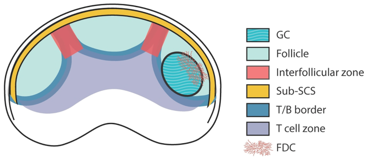
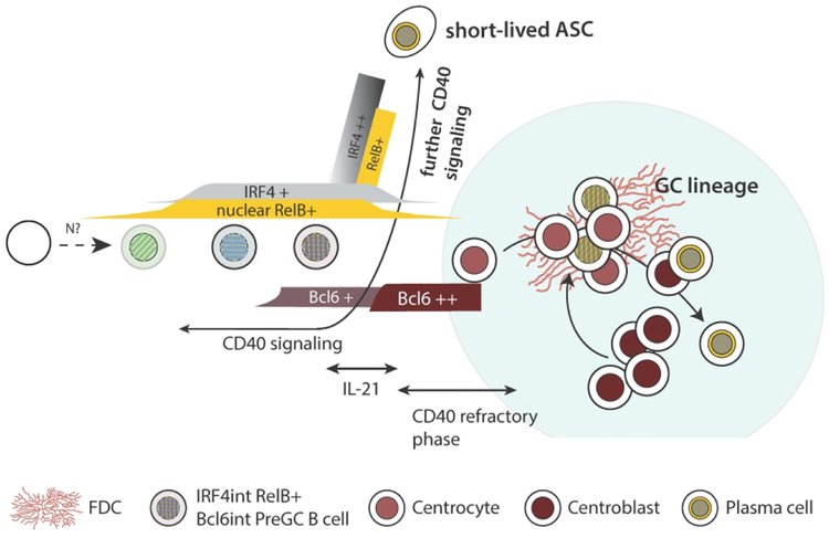
Throughout the developing GC response, B cell survival and fate choices made at the single cell level are dependent on signals received largely through interactions with other cells, often with cognate T cells. The type of signals that a given B cell can encounter is dictated by its location within tissue microarchitecture. The focus of this review is on the initiation and evolution of the GC response at the earliest time points. Here, we review the key factors influencing the progression of GC B cell differentiation that are both stage and context dependent. Finally, we describe the coevolution of niches within and surrounding the GC that influence the outcome of the GC response.

Keywords: B cells; cell differentiation; cytokines; lineage commitment/specification; stromal cells; transcription factors.


Figures

Figure 1.
Figure 1.
Lymph node tissue compartments influencing the developmental stages of germinal center B cells.
Figure 2.
Figure 2.
Temporal and context dependent shifts in transcription factor composition during GC B cell development.
Figure 3.
Figure 3.
Stages of GC maturation and associated tissue niches. (A) Schematic representation of the stages of germinal center (GC) maturation. (B - E) Immunofluorescence staining of popliteal lymph node (B) and spleen (C-E) from C57BL/6 mice that received an adoptive transfer of OVA-specific T cells and antigen-specific B cells enriched by immunomagnetic purification (StemCell Technologies). T cells specific for OVA were obtained from spleens of OT-II mice while either B1-8+ Jκ-/- or MD4 mice served as donors for anti-NP or anti-HEL specific B cells respectively. Recipients were immunized after transfer with 50 μg of either NP-OVA/CFA in the footpad (lymph node, panels B), NP-OVA/alum injected i.p. (spleen, panels C, E), or HEL-OVA/alum (spleen, panel D). (B) Popliteal lymph node histology time-course demonstrating GC maturation after immunization. Stages of maturation represented include the unimmunized follicle (d0/naïve), maturation of the FDC network and light zone expansion (day 4), centroblast increase, zonal segregation and follicle swelling (day 5), and a mature GC (day 10). Sections were stained to highlight CD4+ T cells (blue), IgD+ non-responding B cells within the follicular mantel (green), and CD35+ FDC network (red). (C) Histology showing a maturing GC (day 6) stained to highlight IgD+ naïve B cells (grayscale), PNA+ GC cells (green), and the distribution of sonic hedgehog (SHH, red) in relation to the FDC network stained with CD35 (blue). (D) Representative histology of splenic GCsdemonstrating initial differentiative events shortly after the arrival of Ag-specific B cells to the follicle interior(day 3). Sections were stained to highlight GFP+ Ag-specific B cells (green), CD35 FDCs (blue), Bcl6 (red) toidentify GC committed B cells, and IgMa to assess the level of cytosolic immunoglobulin. Arrows point to Bcells within the FDC network that are cIg hi (consistent with ASC effector lineage), Bcl6hi (GC lineage), andBcl6neg cIg lo (of an undefined alternative fate). (E) Histology showing GFP+ Ag-specific B cell interactionwith dendritic cells (DC) at the T-B border (day 6). Section were stained for CD4+ T cells (blue), B220+ Bcells (red), GFP+ Ag-specific B cells (green) and CD11c+ DC cells (grayscale). Higher magnification imagesof the T-B border demonstrate the ability of GC B cells, DCs and T cells at the T zone border to directly engage each other.

References

    1. Foy TM, Laman JD, Ledbetter JA, Aruffo A, Claassen E and Noelle RJ. gp39-CD40 interactions are essential for germinal center formation and the development of B cell memory. J Exp Med. 1994; 180: 157–63. - PMC - PubMed
    1. Linton PJ, Lo D, Lai L, Thorbecke GJ and Klinman NR. Among naive precursor cell subpopulations only progenitors of memory B cells originate germinal centers. Eur J Immunol. 1992; 22: 1293–7. - PubMed
    1. Linton PL, Decker DJ and Klinman NR. Primary antibody-forming cells and secondary B cells are generated from separate precursor cell subpopulations. Cell. 1989; 59: 1049–59. - PubMed
    1. Tarlinton DM and Smith KG. Dissecting affinity maturation: a model explaining selection of antibody-forming cells and memory B cells in the germinal centre. Immunol Today. 2000; 21: 436–41. - PubMed
    1. MacLennan IC. Germinal centers. Annu Rev Immunol. 1994; 12: 117–39. - PubMed

Publication types