

doi: 10.1038/s41467-019-09231-9.
Automating multimodal microscopy with NanoJ-Fluidics
Affiliations
- PMID: 30874553
- PMCID: PMC6420627
- DOI: 10.1038/s41467-019-09231-9
Item in Clipboard
Automating multimodal microscopy with NanoJ-Fluidics
Nat Commun.
.
Abstract
Combining and multiplexing microscopy approaches is crucial to understand cellular events, but requires elaborate workflows. Here, we present a robust, open-source approach for treating, labelling and imaging live or fixed cells in automated sequences. NanoJ-Fluidics is based on low-cost Lego hardware controlled by ImageJ-based software, making high-content, multimodal imaging easy to implement on any microscope with high reproducibility. We demonstrate its capacity on event-driven, super-resolved live-to-fixed and multiplexed STORM/DNA-PAINT experiments.
Conflict of interest statement
The authors declare no competing interests.
Figures

Schematics of the NanoJ-Fluidics system. a 3D side view of a single syringe pump. b 2D top view of a syringe pump array (representing 4 pumps out of 128 maximum) and a fluid extraction peristaltic pump, both controlled by an Arduino UNO. c Example of possible workflows
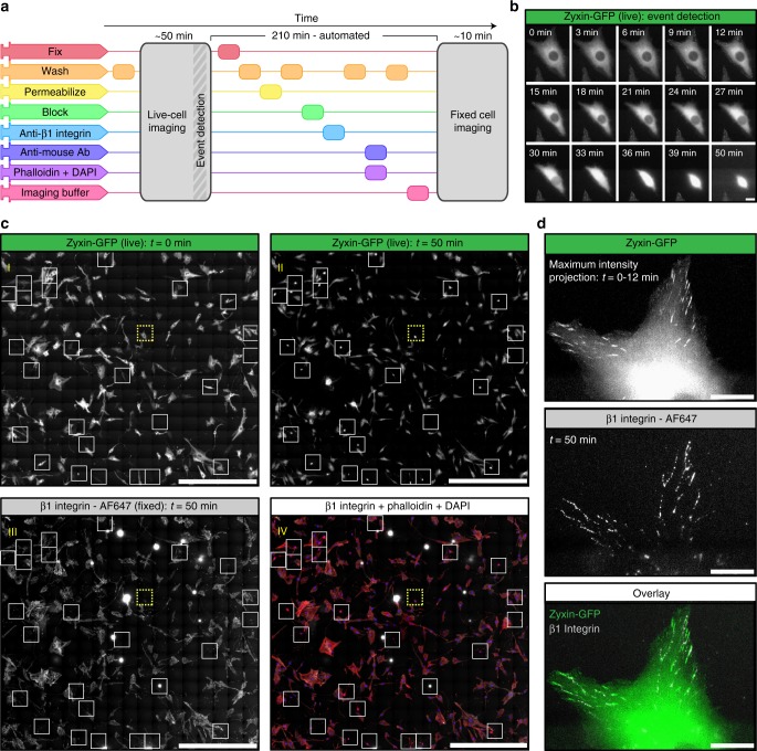
Event-driven fixation of cells upon mitotic rounding. a NanoJ-Fluidics workflow of the event-driven protocol performed. b Stills of RPE1 zyxin-GFP live-cell timelapse during mitotic rounding. Scale bar, 20 μm. c Stitched mosaic (17 × 17 individual regions) of: I—First frame of the live-cell timelapse; II—Last frame of the live-cell timelapse; III—RPE1 zyxin-GFP cells immunolabelled for active β1-integrin; IV—Overlay of RPE1 zyxin-GFP cells immunolabelled for active β1-integrin and stained for F-actin (with phalloidin-TRITC) and DNA (with DAPI). White boxes represent cells where mitotic rounding was observed (Supplementary Movie 4), yellow dashed inset is the cell in (b) and (d). Scale bar is 1 mm. d I—Maximum intensity projection of the first 12 min in (b); II—Active β1-integrin staining; III—Overlay of both panels. Scale bar, 20 μm

Unsupervised live-to-fixed microscopy triggered by mitotic rounding. a NanoJ-Fluidics workflow for the automated mitotic-rounding-driven protocol. b Stills of a HeLa cell expressing mEGFP-α-Tubulin (green) imaged live every 5 min (Supplementary Movie 2) during mitotic cell rounding, corresponding circularity analysis (graph) and overlay of the same cell after the NanoJ-Fluidics protocol (stained for actin—red, phalloidin-AF647 - and DNA—blue, DAPI). Rounding of cell shown was detected at 15 min. Pumps were activated at 35 min, 15 min after 25% of cells reached circularity threshold (Supplementary Note 5). Scale bar, 10 μm. c Two fields-of-view from the same experiment as in panel (b) where mitotic rounding occurred and corresponding single-plane (full z-stack shown in Supplementary Movie 2) of fixed-cell imaging of post NanoJ-Fluidics protocol, overlays and single images coloured as in panel (b). Scale bar, 10 μm
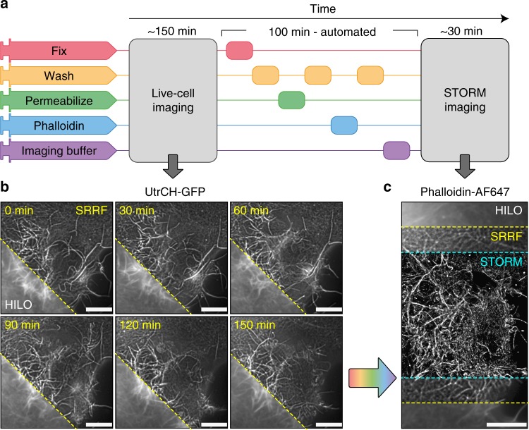
Super-resolution live-to-fixed cell imaging of actin. a NanoJ-Fluidics workflow used for live-to-fixed super-resolution imaging. b HILO and SRRF microscopy images of a COS7 cell expressing UtrCH-GFP imaged every 10 min for 150 min (zoomed in region is shown at 30 min intervals, Supplementary Movie 3 shows extended timelapse and field-of-view). c HILO and SRRF microscopy images of UtrCH-GFP at t = 150 min and the corresponding STORM image after fixation and staining with phalloidin-AF647. Scale bars are 10 μm

Automated DNA-PAINT and STORM imaging. a NanoJ-Fluidics workflow used for STORM and DNA-PAINT imaging. b Left, full view showing 5-channel merge of STORM and DNA-PAINT with actin (yellow, Phalloidin-ATTO488), vimentin (blue, Cy3B DNA-PAINT imager strand), β-tubulin (green, ATTO655 DNA-PAINT imager strand), clathrin (cyan, Cy3B DNA-PAINT imager strand) and mitochondria (red, ATTO655 DNA-PAINT imager strand). Right, zoom of the boxed area. c Single-channel image of each imaged target (top), with insets (bottom) showing a zoom of the boxed area. A movie corresponding to this experiment is available as Supplementary Movie 4. Scale bar corresponds to 10 μm for full images and 2 μm for zooms
References
Publication types
Grants and funding
LinkOut - more resources
Full Text Sources
Other Literature Sources
Research Materials
