. 2019 Mar 15;10(1):1228.
doi: 10.1038/s41467-019-09145-6.

A phosphorylated transcription factor regulates sterol biosynthesis in Fusarium graminearum

Affiliations

A phosphorylated transcription factor regulates sterol biosynthesis in Fusarium graminearum

Zunyong Liu et al. Nat Commun. .

Abstract

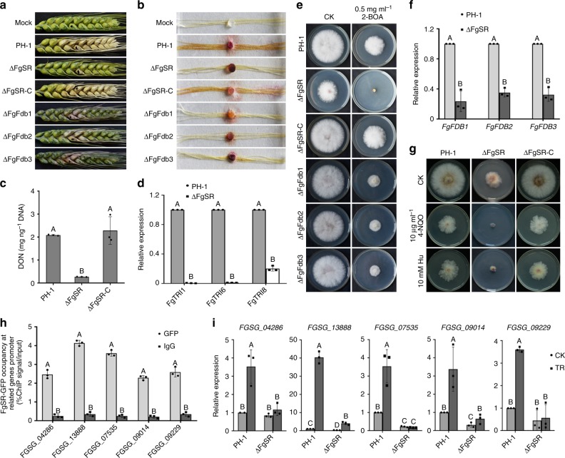
Sterol biosynthesis is controlled by transcription factor SREBP in many eukaryotes. Here, we show that SREBP orthologs are not involved in the regulation of sterol biosynthesis in Fusarium graminearum, a fungal pathogen of cereal crops worldwide. Instead, sterol production is controlled in this organism by a different transcription factor, FgSR, that forms a homodimer and binds to a 16-bp cis-element of its target gene promoters containing two conserved CGAA repeat sequences. FgSR is phosphorylated by the MAP kinase FgHog1, and the phosphorylated FgSR interacts with the chromatin remodeling complex SWI/SNF at the target genes, leading to enhanced transcription. Interestingly, FgSR orthologs exist only in Sordariomycetes and Leotiomycetes fungi. Additionally, FgSR controls virulence mainly via modulating deoxynivalenol biosynthesis and responses to phytoalexin.


Conflict of interest statement

The authors declare no competing interests.

Figures

Fig. 1
Fig. 1
Transcription factor FgSR regulates ergosterol biosynthesis in F. graminearum. a Comparisons in colony morphology among the wild-type PH-1, ΔFgSR and the complemented transformant ΔFgSR-C grown on PDA, CM, or MM. b ΔFgSR displayed increased sensitivity to azole compounds but not to the compounds targeting the high osmolarity glycerol (HOG) pathway. A 5-mm mycelial plug of each strain was inoculated on PDA alone or supplemented with 5 μg ml–1 tridiamenfon, 0.25 μg ml–1 tebuconazole, 10 μg ml–1 iprodione or 0.1 μg ml–1 fludioxonil, and then incubated at 25 °C for 2 days. c Relative abundance of ergosterol in each strain after growth in YEPD for 36 h. Data presented are the mean ± s.d. (n = 3). Bars followed by the same letter are not significantly different according to a Fisher’s least significant difference (LSD) at P = 0.01. d The FgSR amino acid sequence shows a typical Zn(II)2-Cys6 zinc finger domain and a fungal specific transcription factor domain identified using the SMART protein database (http://smart.embl-heidelberg.de) and the NBCI protein database (https://blast.ncbi.nlm.nih.gov/Blast.cgi). e FgSR-GFP localized to the nucleus. Bar = 10 μm
Fig. 2
Fig. 2
FgSR enriches at the promoters of three FgCYP51 genes and regulates their transcription in F. graminearum. a The transcription of FgCYP51A and FgCYP51B was regulated by FgSR, whereas, expression of FgCYP51C was modulated by FgSR only under the ergosterol-absent condition. Each strain was cultured in YEPD for 36 h, and then treated with 2.5 μg ml–1 tebuconazole for 6 h (left panel). ΔFgErg4 was used to mimic the ergosterol-absent condition (right panel). The expression level of each FgCYP51 in PH-1 without treatment was referred to 1 and the FgACTIN gene was used as the internal control for normalization. Data presented are the mean ± s.d. (n = 3). Bars followed by the same letter are not significantly different according to a LSD test at P = 0.01. b FgSR binds to the promoters of three FgCYP51 genes, but the enrichment of FgSR at FgCYP51A and FgCYP51B is more than that at FgCYP51C. Each strain was incubated in YEPD liquid medium for 36 h. ChIP- and input-DNA samples were quantified by quantitative PCR assays with primers amplifying different regions indicated on the diagram of each FgCYP51 promoter. A control reaction was processed in parallel with rabbit IgG and PH-1 transformed only with GFP used as a negative control
Fig. 3
Fig. 3
Phosphorylation of FgSR mediated by FgHog1 regulates the expression of FgCYP51A. a Deletion of kinase in the HOG cascade or mutations at the predicted phosphorylation sites within FgSR led to increased sensitivity to tebuconazole. b Comparisons of expression level of FgCYP51A among the above strains after the treatment with 2.5 μg ml–1 tebuconazole for 6 h. The expression level of FgCYP51A in the wild type without treatment was referred to 1. Data presented are the mean ± s.d. (n = 3). Bars followed by the same letter are not significantly different according to a LSD test at P = 0.01. c The phosphorylation of FgHog1 was increased by the treatment with 2.5 μg ml–1 tebuconazole. PH-1 was treated with tebuconazole for 0, 0.5, 1 and 2 h after incubated in YEPD for 36 h. d Localization of FgHog1-GFP with 2.5 μg ml–1 tebuconazole treatment for 0 and 2 h. Nuclei were stained with 4’,6-diamidino-2-phenylindole (DAPI). DIC, differential interference contrast. Bar = 10 μm. e Loss of FgSsk2, FgPbs2 or FgHog1 decreased the phosphorylation of FgSR with or without tebuconazole treatment. The dephosphorylated and phosphorylated FgSR are indicated with red and blue arrows, respectively. f FgHog1 interacts with FgSR in the co-immunoprecipitation (Co-IP) assay and the interaction is dependent on FgSsk2 and enhanced by tebuconazole treatment. The strains were treated with (+) or without (−) tebuconazole for 2 h after incubated in YEPD for 36 h. g FgHog1 interacts with FgSR in the nucleus under the treatment with 2.5 μg ml–1 tebuconazole, and the interaction is dependent on the FgSsk2 by bimolecular fluorescence complementation (BiFC) assays. Bar = 10 μm. h The interaction of FgHog1 and FgSR assayed using GST pull-downs. Proteins were also detected by staining with Coomassie brilliant blue (CBB). i Phosphorylation of FgSR by Hog1 in vitro. Phosphorylated proteins were resolved by SDS-PAGE and detected by autoradiography (upper panel). GST-FgHog1 and His-FgSR were also detected by staining with CBB (lower panel). j Position of the five predicted phosphorylated amino acid residues in FgSR
Fig. 4
Fig. 4
Phosphorylated FgSR interacts with the SWI/SNF complex to regulate transcription of FgCYP51A. a The yeast two-hybrid (Y2H) assay revealed that the 5 phosphorylation residues of FgSR are critical for its interaction with FgSwp73. b FgSR interacts with FgSwp73 in the co-immunoprecipitation (Co-IP) assay. The relative strains were treated with or without tebuconazole for 2 h after incubated in YEPD for 36 h. c The enrichment of FgSwp73-GFP and FgArp9-GFP at the FgCYP51A promoter among the wild-type PH-1, ΔFgSR, ΔFgSsk2, ΔFgHog1, and ΔFgSR-C5A. ChIP- and input-DNA samples were quantified by quantitative PCR assays with the primer pair A2 indicated in Fig. 2b, and rabbit IgG was used as a control. d Deletion of the SWI/SNF complex subunit FgARP9 led to increased sensitivity to tebuconazole. A 5-mm mycelial plug of PH-1, ΔFgArp9 was inoculated on PDA alone or supplemented with 0.25 μg ml–1 tebuconazole (left panel), and the mycelial growth inhibition was calculated for each strain (right panel). e Comparisons of the expression of FgCYP51A between PH-1 and ΔFgArp9 after the treatment with 2.5 μg ml–1 tebuconazole for 6 h. The expression of FgCYP51A in the wild type without treatment was referred to 1. f The enrichment of histone H3 at the FgCYP51A promoter among the above strains. ChIP- and input-DNA samples were quantified by quantitative PCR assays with the primer pair A2 indicated in Fig. 2b, and rabbit IgG was used as a control. Data presented are the mean ± s.d. (n = 3). Bars followed by the same letter are not significantly different according to a LSD test at P = 0.01
Fig. 5
Fig. 5
The homodimerization of FgSR is required for its binding to the FgCYP51A promoter. a The yeast two-hybrid (Y2H) assay revealed that the middle linker (ML) domain of FgSR is required for its homodimerization. Serial dilutions of yeast cells (cells ml−1) transferred with the prey (pGADT7-FgSR) and bait (a series of truncated FgSR (indicated in the left panel) constructs were assayed for growth on SD-Leu-Trp-His plates (right panel). b FgSR lacking of the ML domain did not interact with itself in the co-immunoprecipitation (Co-IP) assay. c FgSR lacking of the ML domain did not interact with itself in the bimolecular fluorescence complementation (BiFC) assay. FgSR-NYFP/FgSRΔML-NYFP, and FgSR-CYFP/FgSRΔML-CYFP were co-transformed into the wild type. YFP signals were observed using confocal microscopy. Bar = 10 μm. d The strain lacking the zinc finger (ZF) domain (ΔFgSR-CΔZF) and ML domain (ΔFgSR-CΔML) as well as the ΔFgSR mutant displayed greatly increased sensitivity to tebuconazole. e The strains ΔFgSR-CΔZF and ΔFgSR-CΔML exhibited reduced ergosterol content similar to ΔFgSR. f In ΔFgSR-CΔZF and ΔFgSR-CΔML, the expression of FgCYP51A was significantly decreased and could not be induced after the treatment with 2.5 μg ml–1 tebuconazole for 6 h. g FgSRΔML-GFP is still localized in the nucleus. Bar = 10 μm. h ΔFgSR-CΔZF and FgSRΔML-GFP did not enrich at the FgCYP51A promoter. ChIP- and input-DNA samples were quantified by quantitative PCR assays with the primer pair A2 indicated in Fig. 2b, and rabbit IgG was used as a control. Data presented are the mean ± s.d. (n = 3). Bars followed by the same letter are not significantly different according to a LSD test at P = 0.01
Fig. 6
Fig. 6
FgSR is a master regulator of ergosterol biosynthesis in F. graminearum. a ChIP-Seq and -qPCR assays showed that FgSR binds to the promoters of 20 ergosterol biosynthesis genes indicated in blue. RNA-Seq further confirmed that 14 out of the above 20 genes were down-regulated in the ΔFgSR mutant. FgACTIN was use as a negative control in the ChIP-qPCR assay. ChIP- and input-DNA samples were quantified by quantitative PCR assays with the primer pair A2 indicated in Fig. 2b, and rabbit IgG was used as a control. The red arrow indicates the gene direction from 5′ to 3′. The green bar represents the gene promoter region, which is covered during ChIP-Sequencing. “black dashed line” indicates no change in gene transcription determined by the RNA-Seq assay (Supplementary data 3). The sequences below the kurtosis graphs represent the cis-elements within the promoters of the 20 ergosterol pathway genes identified by MEME analysis. Values on the bars followed by the same letter are not significantly different at P = 0.01. b FgSR regulates F. graminearum sensitivity to sterol biosynthesis inhibitors (SBIs) targeted to different biosynthetic enzymes. A 5-mm mycelial plug of each strain was inoculated on PDA supplemented with 5 μg ml–1 naftifine, 0.5 μg ml–1 terbinafine, 5 μg ml–1 fluvastatin, 5 μg ml–1 lovastatin, 10 μg ml–1 spiroxamine, 5 μg ml–1 tridemorph, or 0.25 μg ml–1 prochloraz and then incubated at 25 °C for 3 days. As a control, each strain was cultured on PDA without supplementation
Fig. 7
Fig. 7
Identification of cis-element of FgSR in F. graminearum. a The putative sequence of the cis-element obtained by analyzing the promoters of five target genes with the MEME program. Two CGAA-repeated sequences within the cis-elementare indicated by the black squares. b Schematic representation of modified cis-element in the promoter of FgCYP51A. Two CGAA-repeated sequences are indicated by the black squares. c The cis-element-deleted (ΔFgCYP51A::P1-FgCYP51A) or -mutated (ΔFgCYP51A::P2-FgCYP51A and ΔFgCYP51A::P3-FgCYP51A) strains displayed increased sensitivity to tebuconazole. d In the strains ΔFgCYP51A::P1-FgCYP51A, ΔFgCYP51A::P2-FgCYP51A, and ΔFgCYP51A::P3-FgCYP51A, the expression of FgCYP51A was reduced and could not be induced by the treatment with 2.5 μg ml–1 tebuconazole. The expression level of FgCYP51A in the wild type without treatment was set to 1. e ChIP-qPCR assay revealed that FgSR could not bind to the FgCYP51A promoter lacking the cis-element or containing a mutated cis-element. ChIP- and input-DNA samples were quantified by quantitative PCR assays with the primer pair A2 indicated in Fig. 2b; rabbit IgG was used as a control. Data presented are the mean ± s.d. (n = 3). Bars followed by the same letter are not significantly different according to a LSD test at P = 0.01. f FgSR binds to the cis-element in the FgCYP51A promoter as indicated by yeast one-hybrid (Y1H) assays.The native FgCYP51A promoter (pAbAi-P0), cis-element-deleted and mutated FgCYP51A promoters (pAbAi-P1 to -P3) or the two repeats of the cis-element (pAbAi-P4) was used as a bait, and the pGADT7-FgSR as the prey. g Verification of the binding of FgSR with the cis-element by electrophoretic mobility shift assay (EMSA). Lane 1 and 2, biotin-labeled motif of P0 (as indicated in Fig. 7b) without FgSRN1–158 (lane 1) or with FgSRN1–158 (lane 2); lane 3–5, biotin-labeled motif of P0 with 100 fold excess of unlabelled motif of P0 (lane 3), unlabeled mutated motif of P2 (lane 4), unlabeled mutated motif of P3 (lane 5) with FgSRN1–158; lanes 6 and 7, biotin-labeled mutated motif of P2 and P3 respectively with FgSRN1–158
Fig. 8
Fig. 8
FgSR homologs and their binding cis-element are highly conserved in Sordariomycetes and Leotiomycetes fungi. a FgSR orthologs share high homology in Sordariomycetes and Leotiomycetes fungi, and the FgSR-binding cis-elements within the 1.2-kb promoters of the CYP51 orthologous genes were also detected only in these fungi. The phylogenetic tree was constructed based on the amino acid sequences of RPB2 from 27 eukaryotic species with Mega 5.0 using the neighbor-joining method. The bootstrap values from 1000 replications are indicated on the branches. The homology among FgSR orthologs from 27 species were analyzed using the BLASTMatrix tool on the Comparative Fungal Genomics Platform (http://cfgp.riceblast.snu.ac.kr/). The depth of blue color on the right indicates the level of homology with FgSR. b Deletion of the FgSR ortholog in the Sordariomycetes fungus Fusarium oxysporum led to elevated sensitivity to sterol biosynthesis inhibitor (SBIs). A 5-mm mycelial plug of each strain was inoculated on PDA alone or supplemented with a SBI compound, and then incubated at 25 °C for 2 days. The non-sterol biosynthesis inhibitors, iprodione and pyrimethanil, were used as a control
Fig. 9
Fig. 9
FgSR is required for virulence in F. graminearum. a Flowering wheat heads were point inoculated with a conidial suspension at 105 conidia ml–1 of each strain and infected wheat heads were photographed 15 days after inoculation (dai). b Corn silks were inoculated with mycelial plugs of each strain and examined 4 dai. c The amounts of DON (per ng DNA) produced by the wild type, ΔFgSR and ΔFgSR-C in infected wheat kernels was determined after 20 days of inoculation. d Relative expression levels of DON biosynthetic gene FgTRI1, FgTRI6, and FgTRI8 in wild type and the ΔFgSR mutant grown in TBI for two days. e Sensitivity of PH-1, ΔFgSR, ΔFgSR-C, ΔFgFdb1, −2 and −3 to the phytoalexin 2-benzoxazolinone (BOA). f FgFDB1, FgFDB2 and FgFDB3 were induced in the wild type but not in the ΔFgSR mutant after the treatment with 1 mg ml–1 BOA. The expression level of each gene in the wild type with treatment was set to 1. g Sensitivity of PH-1, ΔFgSR and ΔFgSR-C to DNA damage agent4-nitroquinoline 1-oxide (4-NQO) and hydroxyurea (Hu). h The enrichment of FgSR-GFP at the promoters of five genes related to DNA replication. ChIP- and input-DNA samples were quantified by PCR using primers in each gene promoter. Rabbit IgG was used in as a control. i Relative expression levels of DNA replication-related genes in PH-1 and the ΔFgSR mutant after the treatment with 50 μg ml–1 4-NQO for 4 h. The expression level of each gene in the wild type without treatment was normalized to 1. Data presented are the mean ± s.d. (n = 3). Bars followed by the same letter are not significantly different according to a LSD test at P = 0.01
Fig. 10
Fig. 10
A proposed model for the regulation of sterol biosynthesis mediated by FgSR in F. graminearum. Under the treatment with sterol biosynthesis inhibitors (SBIs), the HOG cascade FgSsk2-FgPbs2-FgHog1 is activated in F. graminearum. Subsequently, FgSR is strongly phosphorylated by activated FgHog1 and other kinase(s). The highly phosphorylated FgSR recruits the SWI/SNF complex to remodel chromatin, and subsequently induces the high transcription levels of sterol biosynthesis genes

Similar articles

Cited by

References

    1. Wollam J, Antebi A. Sterol regulation of metabolism, homeostasis, and development. Annu. Rev. Biochem. 2011;80:885–916. doi: 10.1146/annurev-biochem-081308-165917. - DOI - PMC - PubMed
    1. Daum G, Lees ND, Bard M, Dickson R. Biochemistry, cell biology and molecular biology of lipids of Saccharomyces cerevisiae. Yeast. 1998;14:1471–1510. doi: 10.1002/(SICI)1097-0061(199812)14:16<1471::AID-YEA353>3.0.CO;2-Y. - DOI - PubMed
    1. Espenshade PJ, Hughes AL. Regulation of sterol synthesis in eukaryotes. Annu. Rev. Genet. 2007;41:401–427. doi: 10.1146/annurev.genet.41.110306.130315. - DOI - PubMed
    1. Brown MS, Goldstein JL. The SREBP pathway: regulation of cholesterol metabolism by proteolysis of a membrane-bound transcription factor. Cell. 1997;89:331–340. doi: 10.1016/S0092-8674(00)80213-5. - DOI - PubMed
    1. Zhang YQ, et al. Requirement for ergosterol in V-ATPase function underlies antifungal activity of azole drugs. PLoS Pathog. 2010;6:e1000939. doi: 10.1371/journal.ppat.1000939. - DOI - PMC - PubMed

Publication types

MeSH terms