. 2019 Jun;137(6):901-918.
doi: 10.1007/s00401-019-01976-3. Epub 2019 Mar 14.

Loss of DPP6 in neurodegenerative dementia: a genetic player in the dysfunction of neuronal excitability

Collaborators, Affiliations

Loss of DPP6 in neurodegenerative dementia: a genetic player in the dysfunction of neuronal excitability

Rita Cacace et al. Acta Neuropathol. 2019 Jun.

Abstract

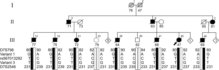
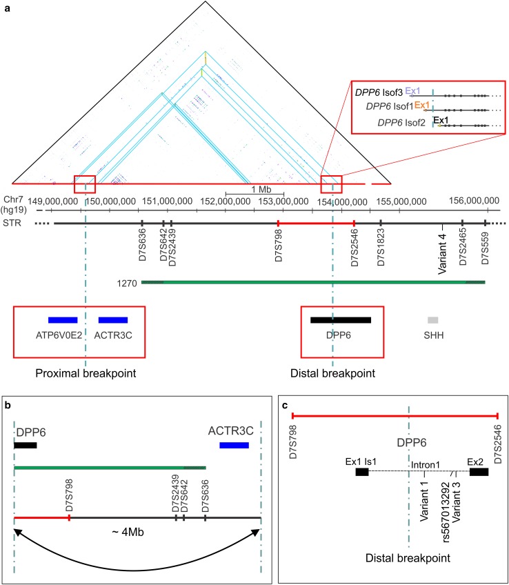
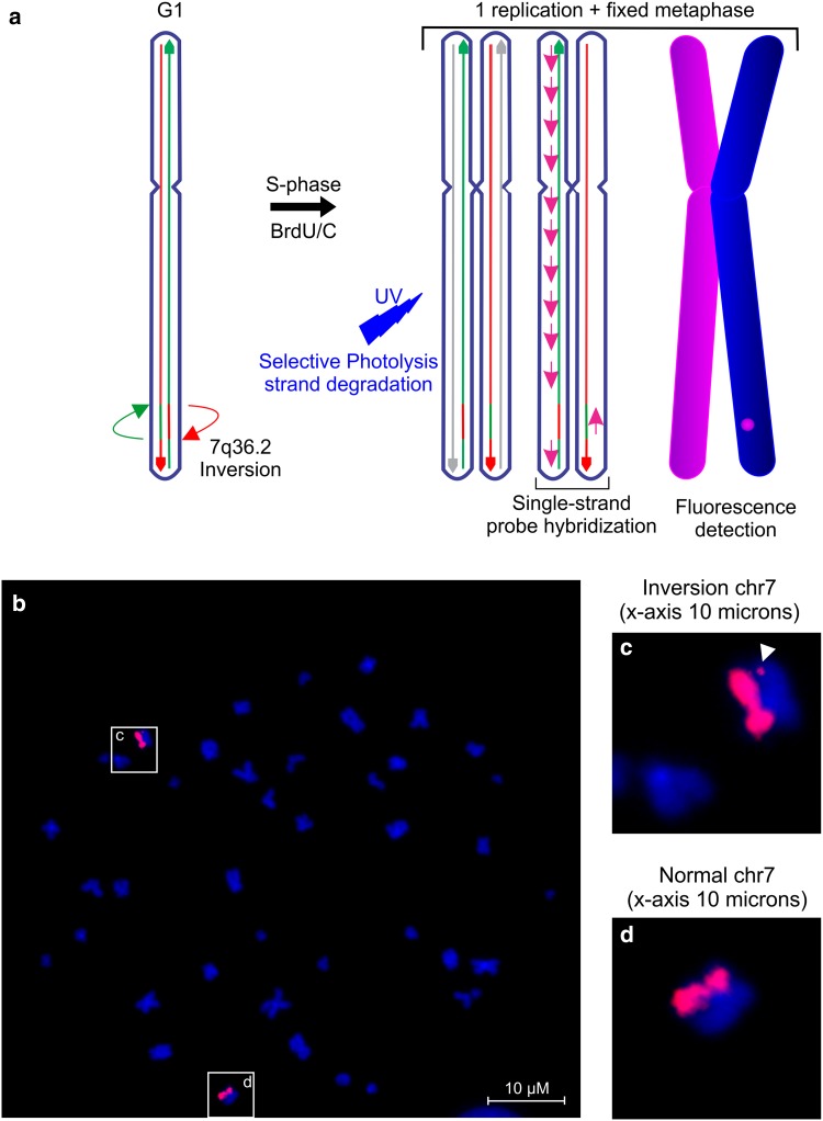
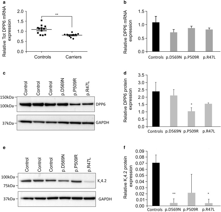
Emerging evidence suggested a converging mechanism in neurodegenerative brain diseases (NBD) involving early neuronal network dysfunctions and alterations in the homeostasis of neuronal firing as culprits of neurodegeneration. In this study, we used paired-end short-read and direct long-read whole genome sequencing to investigate an unresolved autosomal dominant dementia family significantly linked to 7q36. We identified and validated a chromosomal inversion of ca. 4 Mb, segregating on the disease haplotype and disrupting the coding sequence of dipeptidyl-peptidase 6 gene (DPP6). DPP6 resequencing identified significantly more rare variants-nonsense, frameshift, and missense-in early-onset Alzheimer's disease (EOAD, p value = 0.03, OR = 2.21 95% CI 1.05-4.82) and frontotemporal dementia (FTD, p = 0.006, OR = 2.59, 95% CI 1.28-5.49) patient cohorts. DPP6 is a type II transmembrane protein with a highly structured extracellular domain and is mainly expressed in brain, where it binds to the potassium channel Kv4.2 enhancing its expression, regulating its gating properties and controlling the dendritic excitability of hippocampal neurons. Using in vitro modeling, we showed that the missense variants found in patients destabilize DPP6 and reduce its membrane expression (p < 0.001 and p < 0.0001) leading to a loss of protein. Reduced DPP6 and/or Kv4.2 expression was also detected in brain tissue of missense variant carriers. Loss of DPP6 is known to cause neuronal hyperexcitability and behavioral alterations in Dpp6-KO mice. Taken together, the results of our genomic, genetic, expression and modeling analyses, provided direct evidence supporting the involvement of DPP6 loss in dementia. We propose that loss of function variants have a higher penetrance and disease impact, whereas the missense variants have a variable risk contribution to disease that can vary from high to low penetrance. Our findings of DPP6, as novel gene in dementia, strengthen the involvement of neuronal hyperexcitability and alteration in the homeostasis of neuronal firing as a disease mechanism to further investigate.

Keywords: DPP6; Dementia; Hyperexcitability; Neurodegenerative brain diseases; Oxford nanopore technologies (ONT) PromethION; Whole genome sequencing.


Conflict of interest statement

The authors declare no competing financial interests.

Figures

Fig. 1
Fig. 1
Segregation of DPP6 in family 1270. Segregation analysis of three rare variants identified in WGS data in intron 1 of DPP6 (hg18 variant 1 g.153577081 A>G, variant 2 g.153737600 C>T; rs567013292 and variant 3 g.153744958 G>T) delimited by the STR markers D7S798 and D7S2546. Black bars represent the disease haplotype of patients. Numbers within each diamond are unaffected individuals, non-carriers of the disease haplotype included in the genotyping. Arabic numbers above the symbols denote individuals, Arabic numbers below the symbols denote age at onset for patients or either age at last examination or age at death for unaffected individuals. The arrow identifies the proband in the family. WGS data were generated for patients III-12, III-38, III-41, and III-48 from three different sib ships of the pedigree. Direct long-read WGS on Oxford Nanopore PromethION sequencer was performed for III-48. Directional genomic hybridization was performed in cell lines derived from patient III-48 (Fig. 3) and from the non-carriers III-23 and III-39
Fig. 2
Fig. 2
DNA local alignment and schematic representation of 7q36 inversion disrupting DPP6. a DNA local alignment analysis of the NCBI hg19 reference sequence of chromosome 7: 149,169,800–154,794,690 bp shows inverted low copy repeats (LCRs) indicated by blue triangles. The horizontal green bar represents the candidate region of 5.44 Mb (reference build hg19) linked to 7q36 in family 1270, between the short tandem repeats (STRs) markers D7S636 and D7S559 [65]. The dotted vertical lines mark the locations of the proximal and distal inversion breakpoints, with the distal breakpoint in DPP6 (black bar) and the proximal breakpoint in the intergenic region between the genes ATP6V0E2 and ACTR3C (blue bars). Red rectangles magnify the location of the proximal and distal breakpoints. The distal breakpoint is located within the intron 1 of DPP6. Three isoforms are reported in the figure with independent transcription starting sites and regulatory elements (top right red rectangle). b Visualization of the 180° flip of the genomic sequence by the inversion, separating the regulatory region and exon 1 from the coding sequence of DPP6. c Magnification of the region around the distal inversion breakpoint in intron 1 of DPP6 between D7S798 and D7S2546 (red bar) and the location of the three co-segregating rare variants identified by WGS studies. The fourth segregating variant (variant 4) is reported in a downstream of the SHH gene (grey)
Fig. 3
Fig. 3
Inversion validation by directional genomic hybridization. a Schematic presentation of the chromosome 7 directional genomic hybridization (dGH™) assay. b In vitro visualization of the inversion in patient III-48 using a directional genomic hybridization (dGH™) assay. The inverted fragment is observed as a signal switch between the sister chromatids (arrowhead in the magnified image c). No signal is present in the normal chromosome 7 (magnified image d)
Fig. 4
Fig. 4
RNA and protein expression levels of DPP6 missense variants. a Scatter plot of the DPP6 mRNA expression levels in patients (n = 3) compared to control individuals (n = 4). Each circle (patients) or square (control individuals) represents a single measurement; the graph reports the mean ± standard error of the mean (s.e.m.). **p value = 0.0096. b DPP6 mRNA expression level results of three experiments for each of the variants (grey bars) compared to averaged data of four controls (black bar). c Western blot of DPP6 carriers and control individuals and e western blot of Kv4.2 in DPP6 variant carriers and control individuals. d, f Quantification of the expression levels of DPP6 (d) and Kv4.2 (f), obtained from pooling two independent protein preparations within the same western blot experiment. Quantifications are shown for each of the missense variants (grey bars) compared to control individuals (n = 3, black bar). The relative protein expression is reported as average ± standard deviation of three independent protein preparations and quantifications per sample *p < 0.05, **p < 0.005
Fig. 5
Fig. 5
In silico and in vitro modeling of rare variants (MAF < 1%) identified in the screened cohorts. a On scale representation of rare variants (MAF < 1%) detected by DPP6 resequencing. The structural domains [72] are IC, intracellular domain (blue), TM, transmembrane domain (dark green) and EC, extracellular domain including the α/β hydrolase (pink) and the β-propeller domains (turquoise). Seven predicted glycosylation sites are reported as black balls on sticks. DPP6 is a type II transmembrane protein, the N-terminal (NH3+) and the C-terminal (COO) are marked. Variants located in the transmembrane domain (dark green) and extracellular domain are common to all DPP6 isoforms here are represented on the canonical isoform (NM_130797.2). Variants in the intracellular domain (exon 1) are isoform specific. Apart from variants in the variable intracellular domain in the canonical isoform NM_130797.2 (isoform 1), we detected one additional variant (p.A5D) in exon 1 of isoform 3 (NM_001039350.2) not represented in the figure. Represented in red are variants identified in patients only, in green are depicted the variants identified in control individuals only and in black in both patients and control individuals. Variants marked with a black arrow were included in expression studies, since brain tissue of the carriers was available. b Prediction of protein stability in the presence of the missense variants measured in differences in free Gibbs energy (ΔΔG). Destabilizing or stabilizing variants result in positive or negative values, respectively. c In vitro protein stability assay using HiBiT-tagged constructs carrying the variants of interest compared to wild-type DPP6. DPP6 fused to the PEST sequence (WT-PEST) was used as positive control. Graph bars represent normalized luminescence (RLUC) that were used to compare the mutated constructs with the wild-type DPP6. Reported data are the pooled results of six independent experiments, error bars represent standard deviation. ***p < 0.001; ****p < 0.0001

References

    1. Aronesty E. Comparison of sequencing utility programs. Open Bioinform J. 2013;7:1–8. doi: 10.2174/1875036201307010001. - DOI
    1. Auton A, Brooks LD, Durbin RM, Garrison EP, Kang HM, Korbel JO, et al. A global reference for human genetic variation. Nature. 2015;526:68–74. doi: 10.1038/nature15393. - DOI - PMC - PubMed
    1. Baker M, Mackenzie IR, Pickering-Brown SM, Gass J, Rademakers R, Lindholm C, et al. Mutations in progranulin cause tau-negative frontotemporal dementia linked to chromosome 17. Nature. 2006;442:916–919. doi: 10.1038/nature05016. - DOI - PubMed
    1. Bettens K, Brouwers N, Van Miegroet H, Gil A, Engelborghs S, De Deyn PP, et al. Follow-up study of susceptibility loci for Alzheimer’s disease and onset age identified by genome-wide association. J Alzheimers Dis. 2010;19:1169–1175. doi: 10.3233/JAD-2010-1310. - DOI - PubMed
    1. Busche MA, Konnerth A. Impairments of neural circuit function in Alzheimer’s disease. Philos Trans R Soc Lond B Biol Sci. 2016 - PMC - PubMed

Publication types

MeSH terms

Substances