. 2019 Mar 14;11(5):1510-1523.
doi: 10.18632/aging.101849.

Delayed γH2AX foci disappearance in mammary epithelial cells from aged women reveals an age-associated DNA repair defect

Affiliations

Delayed γH2AX foci disappearance in mammary epithelial cells from aged women reveals an age-associated DNA repair defect

Teresa Anglada et al. Aging (Albany NY). .

Abstract

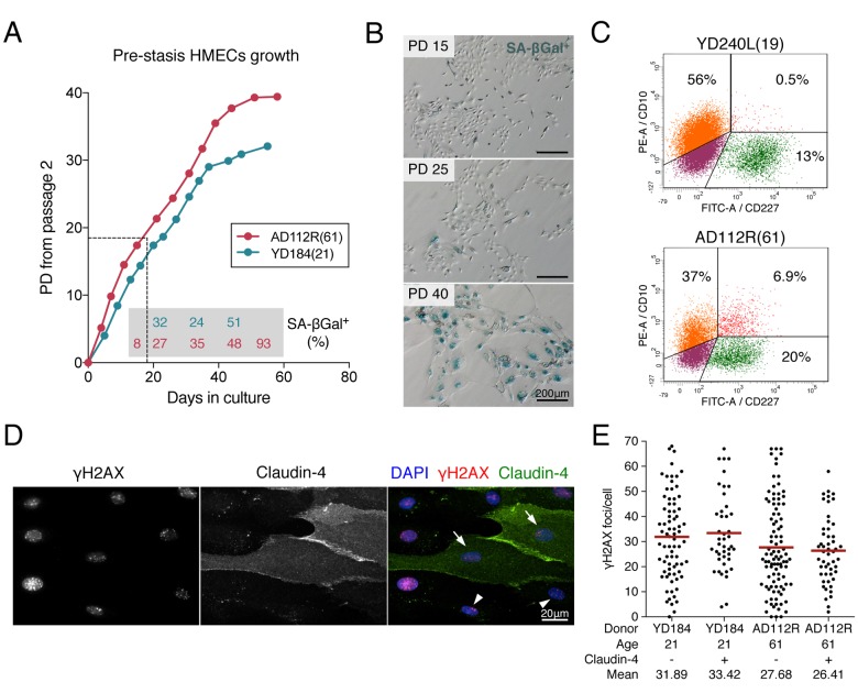
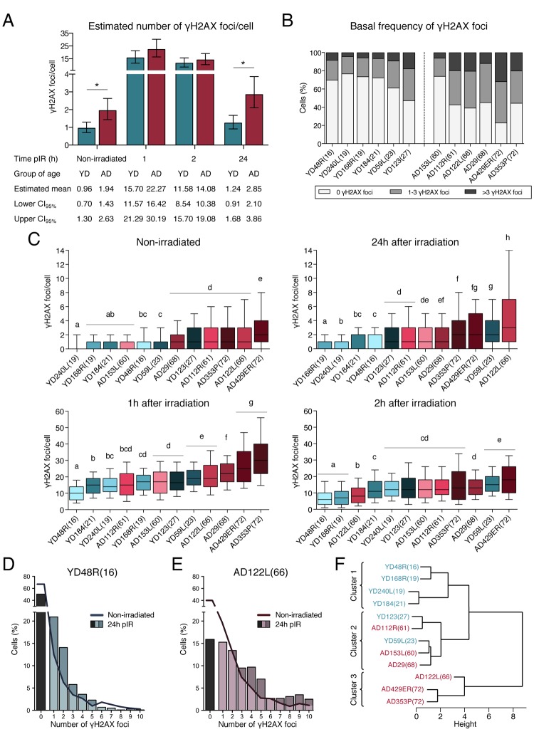
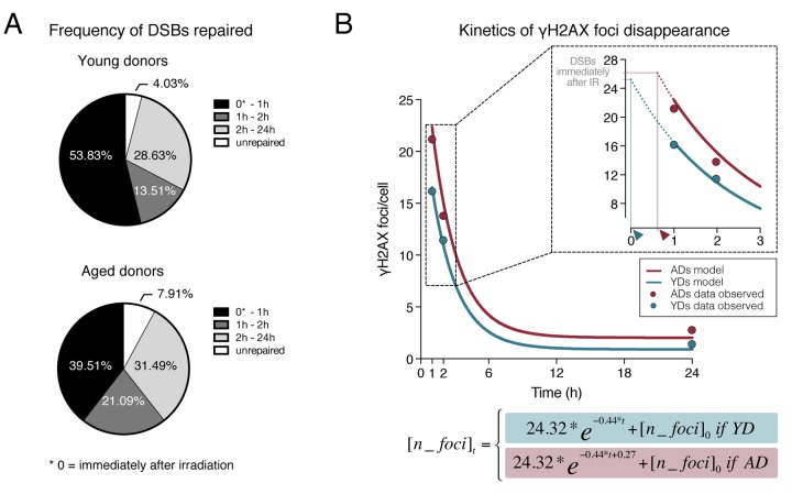
Aging is a degenerative process in which genome instability plays a crucial role. To gain insight into the link between organismal aging and DNA repair capacity, we analyzed DNA double-strand break (DSB) resolution efficiency in human mammary epithelial cells from 12 healthy donors of young and old ages. The frequency of DSBs was measured by quantifying the number of γH2AX foci before and after 1Gy of γ-rays and it was higher in cells from aged donors (ADs) at all times analyzed. At 24 hours after irradiation, ADs retained a significantly higher frequency of residual DSBs than young donors (YDs), which had already reached values close to basal levels. The kinetics of DSB induction and disappearance showed that cells from ADs and YDs repair DSBs with similar speed, although analysis of early times after irradiation indicate that a repair defect may lie within the firing of the DNA repair machinery in AD cells. Indeed, using a mathematical model we calculated a constant factor of delay affecting aged human epithelial cells repair kinetics. This defect manifests with the accumulation of DSBs that might eventually undergo illegitimate repair, thus posing a relevant threat to the maintenance of genome integrity in older individuals.

Keywords: DNA damage; aging; double-strand break repair; genome integrity; human mammary epithelial cells; γH2AX.


Conflict of interest statement

CONFLICTS OF INTEREST: The authors declare that they have no conflicts of interest.

Figures

Figure 1
Figure 1
Pre-stasis HMEC characterization and culture. (A) Representative growth curves of HMECs from YD184(21) and AD112R(61) in M87A medium with supplements. Dots correspond to correlative cell passages from passage 2. The dotted thin line indicates the early passages used for the experiments. Percentages of SA-β-Gal positive cells are indicated within the grey box (N > 500 cells). (B) The frequency of SA-β-Gal positive cells increases with time in culture. (C) Diagrams of flow cytometry analysis of CD10 (PE, phycoerytrin) and CD227 (FITC, fluorescein isothiocyanate) in YD240L(19) and AD112R(61) (N > 10000 cells). (D) Images of the immunofluorescent staining of claudin-4 (expressed by luminal cells, FITC, green), γH2AX (Cy3, red) and DAPI (blue) at 2h after 1Gy of γ-rays exposure. Claudin-4 positive (arrows) and negative (arrowheads) cells are shown. (E) Scatter dot plot and average number (red line) of γH2AX foci/cell in claudin-4 positive and negative cells (N > 100 cells/donor). No statistical differences were observed (Mann-Whitney test, p-value > 0.05).
Figure 2
Figure 2
Frequencies of γH2AX foci in HMECs from young and aged donors. (A) Estimated mean number of γH2AX foci/cell and confidence intervals for young and aged donors. Asterisks indicate significant differences between YDs and ADs (generalized linear model, p-value < 0.01). The number of cells analyzed for each donor is stated in Table 1. (B) Frequency of cells with a defined number of γH2AX foci in non-irradiated samples from the 12 donors. The number of cells analyzed for each donor is stated in Table 1. (C) Box plots of the frequency of γH2AX foci in cells from YDs and ADs in non-irradiated samples and at 1h, 2h or 24h after exposure to 1 Gy of γ-rays. Each donor is colored with blue or red depending on the group of age (blue for YDs and red for ADs). In each group, colors become darker with increasing age of the donor. Boxes include data from the upper to the lower quartile. The median is represented with a black line and whiskers compile 10 to 90% of the scored values. The number of cells analyzed for each donor is stated in Table 1. Statistical differences between donors are indicated following a letter code: donors signaled with the same letter do not show statistical differences and therefore different letters indicate statistically significant differences between donors (Kruskal-Wallis test with Dunn’s multiple comparisons correction, p-value < 0.05). (D, E) Distribution of cells according to the number of γH2AX foci/cell individually scored in YD48R(16) (D) and in AD122L(66) (E). Bars indicate the percentage of cells without foci (black bar) or with ≥1 γH2AX foci (colored bars) 24h after irradiation. The continuous line depicts this percentage before irradiation. The number of cells analyzed for each donor is stated in Table 1. (F) Hierarchical clustering of the 12 donors according to the standardized mean number of γH2AX foci scored in non-irradiated samples and at 1, 2 and 24h after IR. The number of cells analyzed for each donor is stated in Table 1.
Figure 3
Figure 3
Dynamics of γH2AX foci disappearance after irradiation. (A) Frequency of DSBs repaired within defined time intervals after exposure to 1Gy of γ-rays for YDs and ADs. The number of DSBs induced after 1Gy exposure (𝜃) of G1 cells was estimated to be of 35 according to Rothkamm & Löbrich [25]. For the other time points, the numbers of DSBs are those depicted in Tables 1 and 2. (B) Kinetics of γH2AX foci disappearance for young and aged donors following the model of first order kinetic reaction stated in Materials and Methods section. The mean number of γH2AX foci scored at each time point is represented with dots (blue for YDs, red for ADs) and it is stated in Table 1. The lines represent the kinetics of DSBs repair estimated after modeling data of all γH2AX foci/cell from the 12 donors at 1, 2 and 24h after irradiation. The number of cells analyzed for each group of age is stated in Table 1. The inset in the graph shows a detail of the early times after IR exposure. The dotted lines represent an extrapolation of the DSB repair kinetics in the time interval comprised between the DSB repair initiation and 1h after IR. Arrowheads indicate the moment of repair initiation, when the extrapolation lines for YDs and ADs reach the number of γH2AX foci present immediately after IR.

References

    1. López-Otín C, Blasco MA, Partridge L, Serrano M, Kroemer G. The hallmarks of aging. Cell. 2013; 153:1194–217. 10.1016/j.cell.2013.05.039 - DOI - PMC - PubMed
    1. Rogakou EP, Pilch DR, Orr AH, Ivanova VS, Bonner WM. DNA double-stranded breaks induce histone H2AX phosphorylation on serine 139. J Biol Chem. 1998; 273:5858–68. 10.1074/jbc.273.10.5858 - DOI - PubMed
    1. Nakamura AJ, Rao VA, Pommier Y, Bonner WM. The complexity of phosphorylated H2AX foci formation and DNA repair assembly at DNA double-strand breaks. Cell Cycle. 2010; 9:389–97. 10.4161/cc.9.2.10475 - DOI - PMC - PubMed
    1. Rothkamm K, Barnard S, Moquet J, Ellender M, Rana Z, Burdak-Rothkamm S. DNA damage foci: meaning and significance. Environ Mol Mutagen. 2015; 56:491–504. 10.1002/em.21944 - DOI - PubMed
    1. Chowdhury D, Keogh MC, Ishii H, Peterson CL, Buratowski S, Lieberman J. γ-H2AX dephosphorylation by protein phosphatase 2A facilitates DNA double-strand break repair. Mol Cell. 2005; 20:801–09. 10.1016/j.molcel.2005.10.003 - DOI - PubMed

Publication types