. 2019 Mar 14;20(6):1279.
doi: 10.3390/ijms20061279.

Human Heart Explant-Derived Extracellular Vesicles: Characterization and Effects on the In Vitro Recellularization of Decellularized Heart Valves

Affiliations

Human Heart Explant-Derived Extracellular Vesicles: Characterization and Effects on the In Vitro Recellularization of Decellularized Heart Valves

Amanda Leitolis et al. Int J Mol Sci. .

Abstract

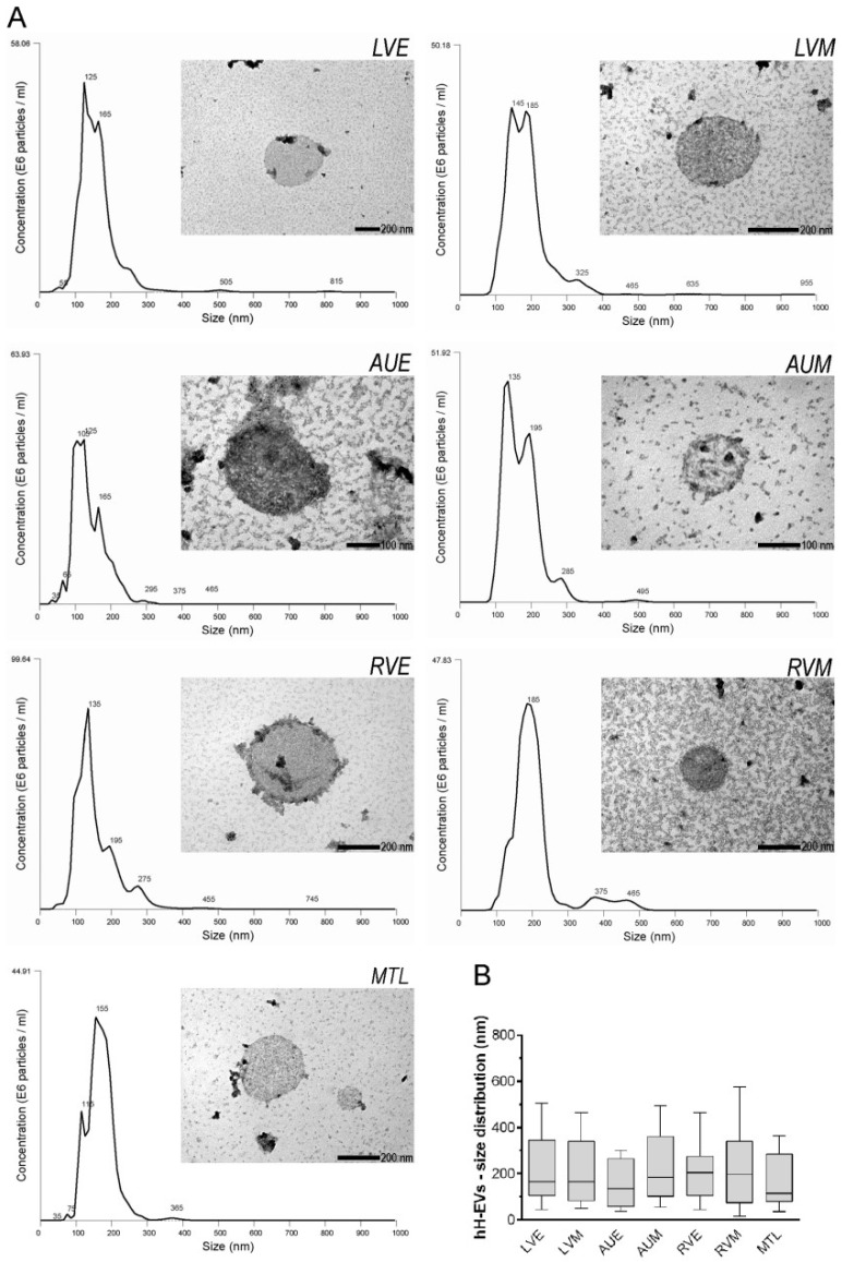
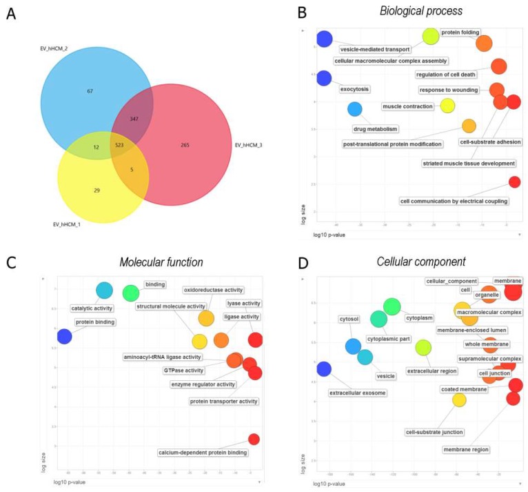
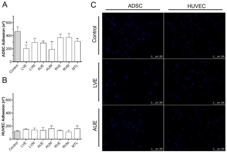
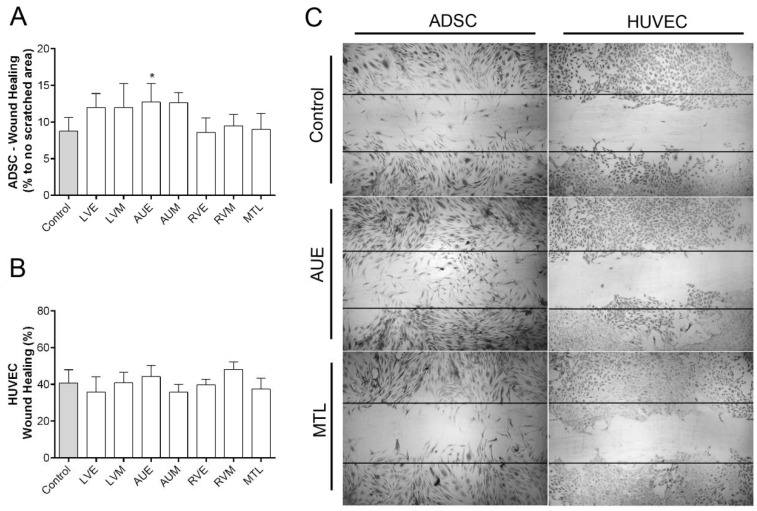
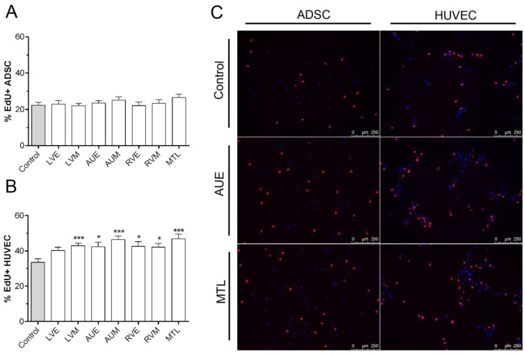
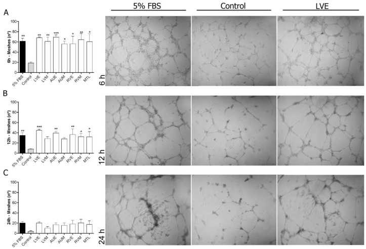
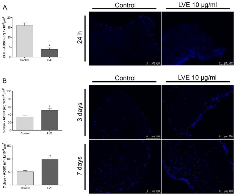
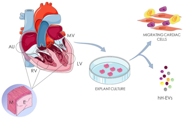
Extracellular vesicles (EVs) are particles released from different cell types and represent key components of paracrine secretion. Accumulating evidence supports the beneficial effects of EVs for tissue regeneration. In this study, discarded human heart tissues were used to isolate human heart-derived extracellular vesicles (hH-EVs). We used nanoparticle tracking analysis (NTA) and transmission electron microscopy (TEM) to physically characterize hH-EVs and mass spectrometry (MS) to profile the protein content in these particles. The MS analysis identified a total of 1248 proteins. Gene ontology (GO) enrichment analysis in hH-EVs revealed the proteins involved in processes, such as the regulation of cell death and response to wounding. The potential of hH-EVs to induce proliferation, adhesion, angiogenesis and wound healing was investigated in vitro. Our findings demonstrate that hH-EVs have the potential to induce proliferation and angiogenesis in endothelial cells, improve wound healing and reduce mesenchymal stem-cell adhesion. Last, we showed that hH-EVs were able to significantly promote mesenchymal stem-cell recellularization of decellularized porcine heart valve leaflets. Altogether our data confirmed that hH-EVs modulate cellular processes, shedding light on the potential of these particles for tissue regeneration and for scaffold recellularization.

Keywords: cardiac regions; extracellular vesicle; heart valve; human heart; mesenchymal stromal cells; recellularization; tissue engineering; tissue explant.


Conflict of interest statement

The authors declare that there is no conflict of interest.

Figures

Figure 1
Figure 1
Characterization of hH-EVs. (A) Representative graphics from nanoparticle tracking analysis (NTA) and transmission electron microscopy (TEM) images of extracellular vesicles derived from cardiac regions: left ventricular endocardium (LVE), left ventricular myocardium (LVM), right auricle endocardium (AUE), right auricle myocardium (AUM), right ventricular endocardium (RVE), right ventricular myocardium (RVM) and mitral valve leaflet (MTL). (B) Analysis of the size and distribution of samples individually examined using NTA.
Figure 2
Figure 2
Protein identification and functional enrichment analysis of human heart-derived extracellular vesicles (hH-EVs). (A) Venn diagram of the proteins identified in three different samples. The diagram shows an overlap of the proteins that were common between the samples analyzed. (BD) Gene ontology enrichment analysis summarized and visualized as a scatter plot using Revigo. The GO terms were ordered in relation to the p-value (x-axis) obtained from the GO term enrichment analysis and the frequency of GO terms in the Gene Ontology Annotation Database (y-axis). The Venn diagram was generated using Funrich software, and the GO analysis was conducted with g:Profiler software.
Figure 3
Figure 3
Influence of hH-EVs derived from cardiac regions on human adipose-derived stem cell (ADSC) and human umbilical vein endothelial cell (HUVEC) adhesion. Quantification of adhered (A) ADSCs and (B) HUVECs stimulated by hH-EVs. (C) Representative images of adhered cells after 20 min of treatment with extracellular vesicles derived from the left ventricular endocardium (LVE) and control without vesicles. Scale bar = 500 µm. * p < 0.05.
Figure 4
Figure 4
Influence of hH-EVs derived from cardiac regions on ADSC and HUVEC wound healing. (A) Quantitative analysis of the percentage of ADSCs in the scratched area after 24 h. (B) Percentage of wound closure by HUVECs after 24 h. (C) Representative images of wound healing stimulated by extracellular vesicles derived from the left ventricular endocardium (LVE) and the right auricle endocardium (AUE). Horizontal lines represent the initial scratched area (0 h), 4× magnification. * p < 0.05.
Figure 5
Figure 5
Influence of hH-EVs derived from cardiac regions on ADSC and HUVEC proliferation. Analysis of the percentage of EdU+ (A) ADSCs and (B) HUVECs cells after 24 h. (C) Representative images of EdU+ cells (red) stimulated by extracellular vesicles derived from right auricle endocardium (AUE) and mitral valve leaflet (MTL). * p < 0.05, *** p < 0.001.
Figure 6
Figure 6
In vitro angiogenesis assay of HUVECs cultured for 24 h on a Matrigel layer under the influence of hH-EVs derived from cardiac regions. Representative images and analysis of the number of meshes formed after 6 h (A), 12 h (B) and 24 h (C). * p < 0.05 vs Control; ** p < 0.01 vs Control; *** p < 0.001 vs Control, 4× magnification.
Figure 7
Figure 7
Extracellular vesicles derived from LVE affect scaffold recellularization. (A) Representative images and analysis of the number of cells/area of fragments previously coated with 10 µg/mL LVE-EVs and cultivated with ADSCs for 24 h. (B) Representative images and analysis of the number of cells/area of fragments cultivated with ADSCs for 24 h and then stimulated with 10 µg/mL LVE-EVs. The fragments were cultivated for 3 and 7 days. Unpaired t test * p < 0.05.
Figure 8
Figure 8
Isolation of heart-derived extracellular vesicles. Human cardiac fragments of right auricle (AU), right ventricle (RV), left ventricle (LV) and mitral valve (MV) were obtained from cadaveric donors. Endocardial (E) and myocardial (M) tissues of AU, RV, LV were separated before culture. The samples were dissociated in fragments of 3 mm² and culture for human heart-derived extracellular vesicles (hH-EVs) and cardiac cells isolation.

References

    1. Gerbin K.A., Murry C.E. The winding road to regenerating the human heart. Cardiovasc. Pathol. 2015;24:133–140. doi: 10.1016/j.carpath.2015.02.004. - DOI - PMC - PubMed
    1. Pinto A.R., Ilinykh A., Ivey M.J., Kuwabara J.T., D’antoni M.L., Debuque R., Chandran A., Wang L., Arora K., Rosenthal N.A., et al. Revisiting cardiac cellular composition. Circ. Res. 2016;118:400–409. doi: 10.1161/CIRCRESAHA.115.307778. - DOI - PMC - PubMed
    1. De Aguiar A.M., Kuligovski C., da Costa M.T.B.A., Stimamiglio M.A., Rebelatto C.L.K., Senegaglia A.C., Brofman P.R.S., Dallagiovanna B., Goldenberg S., Correa A. Alkaline phosphatase-positive cells isolated from human hearts have mesenchymal stem cell characteristics. Stem Cell Discov. 2011;01:71–80. doi: 10.4236/scd.2011.13008. - DOI
    1. Reus T.L., Robert A.W., da Costa M.B.A., de Aguiar A.M., Stimamiglio M.A. Secretome from resident cardiac stromal cells stimulates proliferation, cardiomyogenesis and angiogenesis of progenitor cells. Int. J. Cardiol. 2016;221:396–403. doi: 10.1016/j.ijcard.2016.06.199. - DOI - PubMed
    1. Jackson R., Mount S., Ye B., Mayfield A.E., Chan V., Boodhwani M., Davies R.A., Haddad H., Davis D.R. Isolation of human explant derived cardiac stem cells from cryopreserved heart tissue. PLoS ONE. 2017;12:e0176000. doi: 10.1371/journal.pone.0176000. - DOI - PMC - PubMed

LinkOut - more resources