. 2019 Mar 14;11(3):260.
doi: 10.3390/v11030260.

Enhanced Autophagy Contributes to Reduced Viral Infection in Black Flying Fox Cells

Affiliations

Enhanced Autophagy Contributes to Reduced Viral Infection in Black Flying Fox Cells

Eric D Laing et al. Viruses. .

Abstract

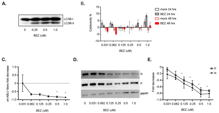
Bats are increasingly implicated as hosts of highly pathogenic viruses. The underlying virus⁻host interactions and cellular mechanisms that promote co-existence remain ill-defined, but physiological traits such as flight and longevity are proposed to drive these adaptations. Autophagy is a cellular homeostatic process that regulates ageing, metabolism, and intrinsic immune defense. We quantified basal and stimulated autophagic responses in black flying fox cells, and demonstrated that although black flying fox cells are susceptible to Australian bat lyssavirus (ABLV) infection, viral replication is dampened in these bat cells. Black flying fox cells tolerated prolonged ABLV infection with less cell death relative to comparable human cells, suggesting post-entry mechanisms interference with virus replication. An elevated basal autophagic level was observed and autophagy was induced in response to high virus doses. Pharmacological stimulation of the autophagy pathway reduced virus replication, indicating autophagy acts as an anti-viral mechanism. Enhancement of basal and virus-induced autophagy in bat cells connects related reports that long-lived species possess homeostatic processes that dampen oxidative stress and macromolecule damage. Exemplifying the potential that evolved cellular homeostatic adaptations like autophagy may secondarily act as anti-viral mechanisms, enabling bats to serve as natural hosts to an assortment of pathogenic viruses. Furthermore, our data suggest autophagy-inducing drugs may provide a novel therapeutic strategy for combating lyssavirus infection.

Keywords: autophagy; bats; viruses.


Conflict of interest statement

The authors declare no conflict of interest. The funders had no role in the design of the study; in the collection, analyses, or interpretation of data; in the writing of the manuscript, or in the decision to publish the results.

Figures

Figure 1
Figure 1
ABLV replication is reduced in black flying fox cells. (A) wt-ABLV and rABLV-GFP genome schematics. (B) rABLV-GFP titers. Cells were incubated with rABLV-GFP (MOI 1) then after two hours the virus inoculated cell culture supernatant was replaced with fresh cell culture media. Cell culture supernatant and cells were collected after at indicated hpi and serial dilutions were incubated with fresh HEK293T cells to determine titers. (C) Flow cytometry analysis of GFP positive cells over a time course of rABLV-GFP infection. (D) Western blot image of ABLV N, P, and vGFP expression in all cell lines at 72 hpi. Data are a representation of two independent experiments, mean ± SEM.
Figure 2
Figure 2
ABLV infection induces less cell death in black flying fox cells. (A) Histogram overlays LIVE/DEAD Violet stained NBF-L and PaBrH cells after rABLV-GFP infection. Percentages of Violet+ dead cells are demarcated for hpi and MOI indicated. (B) Percentage of dead cells expressed as a fold change compared to MOI 0 at each time point. Data are a representative of three independent experiments, * p < 0.05, ANOVA (two-way), and Tukey’s multiple comparison test. (C) Percentage of GFP positive cells at 48 and 96 hpi. (D) Median fluorescence intensity (MFI) of ABLV expressed GFP. Data (CD) are representative of two independent experiments, mean ± SEM, * p < 0.01, ANOVA (two-way), and Tukey’s multiple comparison test.
Figure 3
Figure 3
ABLV infection activates autophagy. (A) LC3B western blot of lysates from human NBF-L and black flying fox PaBrH and PaKiT cells, treated with BAFA1 (400 nM; 2 h). (B) LC3B-II (β-actin-normalized) from BAFA1 treated cells. (C) LC3B western blot of lysates from PaBrH cell lysates. PaBrH cells were infected for times (hpi) and MOI indicated. (D) LC3B-II (β-actin-normalized) expressed as a percentage of total LC3B from PaBrH cells infected with rABLV-GFP. (E) LC3B western blot of lysates from NBF-L cells infected with rABLV-GFP. (F) LC3B-II (gapdh-normalized) from NBF-L cells infected with rABLV-GFP. PaBrH and NBF-L cells were infected with rABLV-GFP for times (hpi) and MOI indicated. All data are a representative of three independent experiments, LC3B-II is expressed as a percentage of total LC3B, mean ± SEM, * p < 0.05, student’s t-test.
Figure 4
Figure 4
ABLV infection and autophagy activation in primary black flying fox brain cells. (A) Western blot for LC3B in primary black flying fox brain (PaBr) cells following 24 and 48 hpi with wt-ABLV MOI indicated. BAFA1 treatment (400 nM) was used as a control for blocking autophagic flux. (B) Quantification of LC3B-II levels (β-actin normalized) relative light units (RLU) from cells infected in (A). All data are representative of three independent experiments, mean ± SEM and * p < 0.05 ANOVA (two-way), and Tukey’s multiple comparison test (48 hpi MOI 0 vs. MOI 10).
Figure 5
Figure 5
ABLV infection does not inhibit autophagic flux. (A) Western blot for p62 and NDP52 proteins in PaBrH cell lysates, rABLV-GFP (MOI 1.0), and BAFA1 (400 nM; 2 h). (B) Log transformed fold change of p62 protein (PaBrH) normalized to β-actin and compared to uninfected DMSO-mock treated time point controls. (C) Log transformed fold change in NDP52 protein (PaBrH) normalized to β-actin and compared to uninfected DMSO-mock treated time point controls. (D) Western blot for p62 in NBF-L cell lysates. (E) Log transformed fold change in p62 (NBF-L) normalized β-actin and compared to uninfected DMSO-mock treated time point controls. All data are representative of three independent experiments, mean ± SEM, * ANOVA (two-way), and Tukey’s multiple comparison test.
Figure 6
Figure 6
Pharmacologic activation of autophagy limits ABLV replication. (A) Flow cytometry histogram overlays of PaKiT cells stained with CYTO-ID®. PaKiT cells were treated with RAPA (2 µM), and SMER (50 µM) for h indicated; as well as CHQ (1 µM, 18 h). (B) Fold change in rABLV-GFP titers (pfu/mL) fold change from PaBrH, PaKiT, and NBF-L cell culture supernatants collected 48 hpi (MOI 1.0). Cells were treated with RAPA (2 µM) and SMER (50 µM) for 24 h. (C) Western blot of ABLV N, P, and vGFP in PaBrH cell lysates. (D) Fold change in ABLV N, P, and vGFP proteins (β-actin-normalized) from PaBrH cell lysates. (E) Western blot of ABLV N, P, and vGFP collected in PaKiT cell lysates. (F) Fold change in ABLV N, P, and vGFP proteins (β-actin-normalized) collected from PaKiT cell lysates. (G) Western blot of ABLV N, P, and vGFP collected in NBF-L cell lysates. (H) Fold change in ABLV N, P, and vGFP proteins (β-actin-normalized) collected from NBF-L cell lysates. (CH) Whole cell lysates were collected 48 hpi (rABLV-GFP MOI 1.0). Fold change is a representation of at least three independent experiments, mean ± SEM and ** p < 0.001, * p < 0.05 ANOVA (one-way) analysis, Dunnett’s multiple comparison test with treatments compared to DMSO mock treated cells.
Figure 7
Figure 7
BEZ treatment reduced wt-ABLV replication in a human neuroblastoma cell line. (A) Western blot of LC3B protein in lysates from NBF-L cells were treated with indicated concentrations of BEZ for 4 h. (B) Lactate dehydrogenase cytotoxicity assay of NBF-L cells treated with BEZ or mock-treated with DMSO. ANOVA analysis revealed no significant differences of quantified spontaneous cytotoxicity between DMSO or BEZ treatments. (C) Fold decreases in wt-ABLV titers (pfu/mL) from cells treated in (A) fold decreases. (D) Western blot image of wt-ABLV N and P protein in NBF-L lysates prepared in (A). (E) Fold decreases in wt-ABLV N and P protein from cells treated in (A). Data are a representative of three independent experiments, mean ± SEM, * p < 0.05 ANOVA (one-way) analysis BEZ (µM) compared to DMSO (mock) treatment.

References

    1. Olival K.J., Hosseini P.R., Zambrana-Torrelio C., Ross N., Bogich T.L., Daszak P. Host and viral traits predict zoonotic spillover from mammals. Nature. 2017;546:646–650. doi: 10.1038/nature22975. - DOI - PMC - PubMed
    1. Halpin K., Hyatt A.D., Fogarty R., Middleton D., Bingham J., Epstein J.H., Rahman S.A., Hughes T., Smith C., Field H.E., et al. Pteropid bats are confirmed as the reservoir hosts of henipaviruses: A comprehensive experimental study of virus transmission. Am. J. Trop. Med. Hyg. 2011;85:946–951. doi: 10.4269/ajtmh.2011.10-0567. - DOI - PMC - PubMed
    1. Towner J.S., Amman B.R., Sealy T.K., Carroll S.A., Comer J.A., Kemp A., Swanepoel R., Paddock C.D., Balinandi S., Khristova M.L., et al. Isolation of genetically diverse marburg viruses from egyptian fruit bats. PLoS Pathog. 2009;5:e1000536. doi: 10.1371/journal.ppat.1000536. - DOI - PMC - PubMed
    1. Amman B.R., Jones M.E., Sealy T.K., Uebelhoer L.S., Schuh A.J., Bird B.H., Coleman-McCray J.D., Martin B.E., Nichol S.T., Towner J.S. Oral shedding of marburg virus in experimentally infected egyptian fruit bats (rousettus aegyptiacus) J. Wildl. Dis. 2015;51:113–124. doi: 10.7589/2014-08-198. - DOI - PMC - PubMed
    1. Li W., Shi Z., Yu M., Ren W., Smith C., Epstein J.H., Wang H., Crameri G., Hu Z., Zhang H., et al. Bats are natural reservoirs of sars-like coronaviruses. Science. 2005;310:676–679. doi: 10.1126/science.1118391. - DOI - PubMed

Publication types

LinkOut - more resources