. 2019 Mar 14;8(3):243.
doi: 10.3390/cells8030243.

Membrane-Associated, Not Cytoplasmic or Nuclear, FGFR1 Induces Neuronal Differentiation

Affiliations

Membrane-Associated, Not Cytoplasmic or Nuclear, FGFR1 Induces Neuronal Differentiation

Katalin Csanaky et al. Cells. .

Abstract

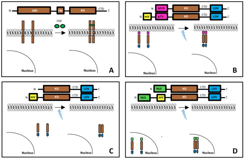
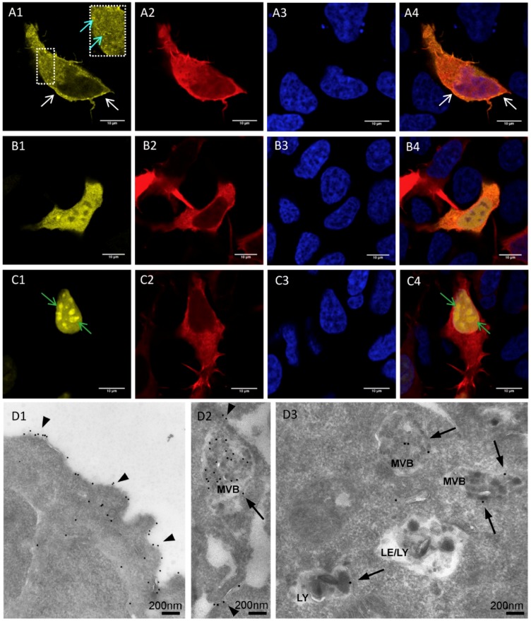
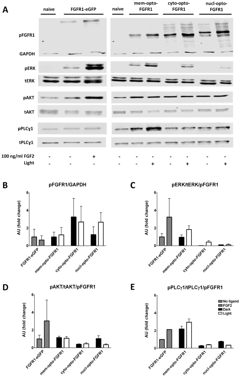
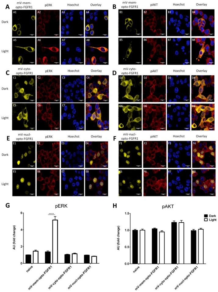
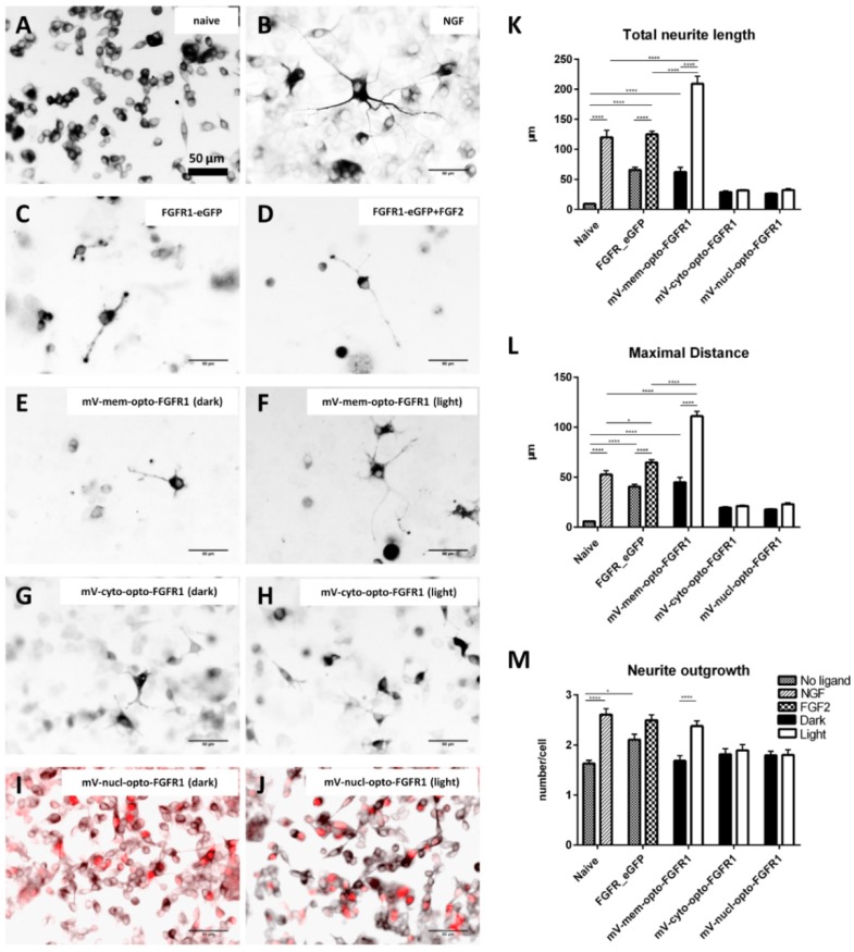
The intracellular transport of receptor tyrosine kinases results in the differential activation of various signaling pathways. In this study, optogenetic stimulation of fibroblast growth factor receptor type 1 (FGFR1) was performed to study the effects of subcellular targeting of receptor kinases on signaling and neurite outgrowth. The catalytic domain of FGFR1 fused to the algal light-oxygen-voltage-sensing (LOV) domain was directed to different cellular compartments (plasma membrane, cytoplasm and nucleus) in human embryonic kidney (HEK293) and pheochromocytoma (PC12) cells. Blue light stimulation elevated the pERK and pPLCγ1 levels in membrane-opto-FGFR1-transfected cells similarly to ligand-induced receptor activation; however, no changes in pAKT levels were observed. PC12 cells transfected with membrane-opto-FGFR1 exhibited significantly longer neurites after light stimulation than after growth factor treatment, and significantly more neurites extended from their cell bodies. The activation of cytoplasmic FGFR1 kinase enhanced ERK signaling in HEK293 cells but not in PC12 cells and did not induce neuronal differentiation. The stimulation of FGFR1 kinase in the nucleus also did not result in signaling changes or neurite outgrowth. We conclude that FGFR1 kinase needs to be associated with membranes to induce the differentiation of PC12 cells mainly via ERK activation.

Keywords: AKT; ERK; FGF2; HEK293; PC12; neurite outgrowth; optogenetics; receptor kinase.


Conflict of interest statement

The authors declare no conflict of interest.

Figures

Figure 1
Figure 1
Design of light-controlled opto-fibroblast growth factor receptors (FGFR1s) and their molecular architecture: sequence of the different FGFR1 coding genes, spatial relation of FGFR1s to the surface membrane and nucleus, and their activation/dimerization. (A) Naturally occurring full-length FGFR1 consists of the extracellular ligand-binding (LBD), transmembrane (TM), kinase (KD) and C-terminal tail (CTD) domains. (B) Artificial mem-opto-FGFR1 is anchored to the plasma membrane with an N-terminal myristoylation signal (MYR) followed by the KD, CTD and LOV domain (mV-mem-opto-FGFR1 is inserted into the plasma membrane by incorporation of the transmembrane domain of p75). Fluorescent mVenus and the light-oxygen-voltage-sensing (LOV) protein are separated to avoid non-specific activation of the LOV domain by mVenus (by Förster resonance energy transfer). mVenus can be detected by excitation with green light (514 nm) that does not activate the LOV domain. (C) Cyto-opto-FGFR1 consists of only the KD and the LOV domain. (D) Three nuclear localization sequence (NLS) signals are inserted into nucl-opto-FGFR1 with or without mVenus.
Figure 2
Figure 2
Light and electron microscopic localization of mV-opto-FGFR1s in human embryonic kidney (HEK293) cells. (AC) Immunofluorescence microscopy of cells co-transfected with mV-opto-FGFR1s and LifeAct–mCherry to visualize cell bodies and all cytoplasmic processes. (A1) mV-mem-opto-FGFR1 is observed in the plasma membrane (white arrows) and in endosomes (inset; cyan arrows) indicating endosomal receptor internalization. (B1) mV-cyto-opto-FGFR1-transfected cells reveal diffuse yellow fluorescence in the cytoplasm and nucleus of transfected cells. (C1) mV-nucl-opto-FGFR1 is only located in the nucleus (nuclear speckle domains are indicated by green arrows). Fixed cell nuclei are stained with Hoechst (blue) and A4–C4 represent overlays. Scale bars for all images are 10 µm. (D) Immunogold electron microscopy of the thawed cryosections reveals mV-mem-opto-FGFR1 in the plasma membrane (arrowheads in D1 and D2) and in the limiting membrane as well as inside various endocytic compartments (arrows in D2 and D3); MVB = multivesicular body, LE = late endosome, LY = lysosome.
Figure 3
Figure 3
Immunoblot analysis of light- and FGF2-induced activation of key signaling molecules in HEK293 cells. (A) Representative examples of Western blots for pFGFR1/GAPDH, pERK/tERK, pAKT/tAKT and pPLCɣ1/tPLCɣ1 are shown. (BE) Average intensities of ERK, AKT, and PLCɣ1 phosphorylation were quantified after normalization for pFGFR1/GAPDH to control for differences in plasmid expression levels. All ratios are relative to FGFR1–eGFP (=1) and calculated from three independent experiments (mean ± SEM).
Figure 4
Figure 4
Light-induced ERK and AKT activation in mV-opto-FGFR1-transfected PC12 cells. Although mV-mem-opto-FGFR1-transfected cells exhibit short neurite extensions (sprouts) in the dark (A1), blue light induces neuronal differentiation with long, slender neurites in transfected cells (A5). (A) Significantly increased cytoplasmic pERK levels following light stimulation of mV-mem-opto-FGFR1-transfected cells, while non-transfected cells show no changes. (C,E) The pERK level is unchanged after stimulation of mV-cyto- and nucl-opto-FGFR1-transfected cells. (B,D,F) The fluorescence intensities of pAKT signals are similar in mV-opto-FGFR1-transfected and non-transfected cells in the dark and no changes are observed after blue light stimulation. Images are acquired using the same laser intensity for both dark and light in each fluorescent channel and presented without adjusting contrast or subtracting background. (G,H) Quantification of average fluorescence intensities. Results are calculated from two independent experiments and presented as mean ± SEM (30 < n < 60), **** p < 0.0001. Scale bars = 10 μm.
Figure 5
Figure 5
Ligand- and light-induced neurite outgrowth by pheochromocytoma (PC12) cells. (AJ) Inverted immunofluorescence images following neuron-specific class III β-tubulin staining to identify neurites (red nuclei in nucl-opto-FGFR1 cells allow identification of transfected cells in I/J). (KM) Quantification of morphological parameters (total neurite outgrowth, longest process and number of processes per cell; see Figure S1 for details). Results are calculated from three independent experiments and presented as mean ± SEM (50 < n < 100), * p < 0.05, **** p < 0.0001. Scale bars = 50 μm.

Similar articles

Cited by

References

    1. Lee P.L., Johnson D.E., Cousens L.S., Fried V.A., Williams L.T. Purification and complementary DNA cloning of a receptor for basic fibroblast growth factor. Science. 1989;245:57–60. doi: 10.1126/science.2544996. - DOI - PubMed
    1. Zhou F.Q., Snider W.D. Intracellular control of developmental and regenerative axon growth. Philos. Trans. R. Soc. Lond. B. Biol. Sci. 2006;361:1575–1592. doi: 10.1098/rstb.2006.1882. - DOI - PMC - PubMed
    1. Eswarakumar V.P., Lax I., Schlessinger J. Cellular signaling by fibroblast growth factor receptors. Cytokine Growth Factor Rev. 2005;16:139–149. doi: 10.1016/j.cytogfr.2005.01.001. - DOI - PubMed
    1. Platta H.W., Stenmark H. Endocytosis and signaling. Curr. Opin. Cell Biol. 2011;23:393–403. doi: 10.1016/j.ceb.2011.03.008. - DOI - PubMed
    1. MacInnis B.L., Campenot R.B. Retrograde support of neuronal survival without retrograde transport of nerve growth factor. Science. 2002;295:1536–1539. doi: 10.1126/science.1064913. - DOI - PubMed

Publication types

MeSH terms

Substances

LinkOut - more resources