Intranasal Borna Disease Virus (BoDV-1) Infection: Insights into Initial Steps and Potential Contagiosity
- PMID: 30875911
- PMCID: PMC6470550
- DOI: 10.3390/ijms20061318
Intranasal Borna Disease Virus (BoDV-1) Infection: Insights into Initial Steps and Potential Contagiosity
Abstract
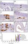
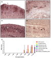
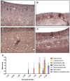
Mammalian Bornavirus (BoDV-1) typically causes a fatal neurologic disorder in horses and sheep, and was recently shown to cause fatal encephalitis in humans with and without transplant reception. It has been suggested that BoDV-1 enters the central nervous system (CNS) via the olfactory pathway. However, (I) susceptible cell types that replicate the virus for successful spread, and (II) the role of olfactory ensheathing cells (OECs), remained unclear. To address this, we studied the intranasal infection of adult rats with BoDV-1 in vivo and in vitro, using olfactory mucosal (OM) cell cultures and the cultures of purified OECs. Strikingly, in vitro and in vivo, viral antigen and mRNA were present from four days post infection (dpi) onwards in the olfactory receptor neurons (ORNs), but also in all other cell types of the OM, and constantly in the OECs. In contrast, in vivo, BoDV-1 genomic RNA was only detectable in adult and juvenile ORNs, nerve fibers, and in OECs from 7 dpi on. In vitro, the rate of infection of OECs was significantly higher than that of the OM cells, pointing to a crucial role of OECs for infection via the olfactory pathway. Thus, this study provides important insights into the transmission of neurotropic viral infections with a zoonotic potential.
Keywords: OECs; borna disease virus; in vitro; in vivo; initial phase; olfactory ensheathing cells; olfactory epithelium.
Conflict of interest statement
The authors declare no conflict of interests.
Figures


Similar articles
-
Zoonotic spillover infections with Borna disease virus 1 leading to fatal human encephalitis, 1999-2019: an epidemiological investigation.Lancet Infect Dis. 2020 Apr;20(4):467-477. doi: 10.1016/S1473-3099(19)30546-8. Epub 2020 Jan 7. Lancet Infect Dis. 2020. PMID: 31924550
-
Generation of a non-transmissive Borna disease virus vector lacking both matrix and glycoprotein genes.Microbiol Immunol. 2017 Sep;61(9):380-386. doi: 10.1111/1348-0421.12505. Microbiol Immunol. 2017. PMID: 28776750
-
Splicing-Dependent Subcellular Targeting of Borna Disease Virus Nucleoprotein Isoforms.J Virol. 2019 Feb 19;93(5):e01621-18. doi: 10.1128/JVI.01621-18. Print 2019 Mar 1. J Virol. 2019. PMID: 30541858 Free PMC article.
-
The potential therapeutic applications of olfactory ensheathing cells in regenerative medicine.Cell Transplant. 2014;23(4-5):567-71. doi: 10.3727/096368914X678508. Cell Transplant. 2014. PMID: 24816451 Review.
-
Reverse genetics approaches of Borna disease virus: applications in development of viral vectors and preventive vaccines.Curr Opin Virol. 2020 Oct;44:42-48. doi: 10.1016/j.coviro.2020.05.011. Epub 2020 Jul 10. Curr Opin Virol. 2020. PMID: 32659515 Review.
Cited by
-
The unfolding palette of COVID-19 multisystemic syndrome and its neurological manifestations.Brain Behav Immun Health. 2021 Jul;14:100251. doi: 10.1016/j.bbih.2021.100251. Epub 2021 Apr 3. Brain Behav Immun Health. 2021. PMID: 33842898 Free PMC article. Review.
-
Comparative study of virus and lymphocyte distribution with clinical data suggests early high dose immunosuppression as potential key factor for the therapy of patients with BoDV-1 infection.Emerg Microbes Infect. 2024 Dec;13(1):2350168. doi: 10.1080/22221751.2024.2350168. Epub 2024 May 20. Emerg Microbes Infect. 2024. PMID: 38687703 Free PMC article.
-
Lethal Borna disease virus 1 infections of humans and animals - in-depth molecular epidemiology and phylogeography.Nat Commun. 2024 Sep 10;15(1):7908. doi: 10.1038/s41467-024-52192-x. Nat Commun. 2024. PMID: 39256401 Free PMC article.
-
Development of a nonhuman primate model for mammalian bornavirus infection.PNAS Nexus. 2022 Jun 8;1(3):pgac073. doi: 10.1093/pnasnexus/pgac073. eCollection 2022 Jul. PNAS Nexus. 2022. PMID: 35860599 Free PMC article.
-
Developing a universal multi-epitope protein vaccine candidate for enhanced borna virus pandemic preparedness.Front Immunol. 2024 Dec 5;15:1427677. doi: 10.3389/fimmu.2024.1427677. eCollection 2024. Front Immunol. 2024. PMID: 39703502 Free PMC article.
References
-
- Herden C., Briese T., Lipkin W.I., Richt J.A. Bornaviridae. In: Knipe D.M., Howley P.M., editors. Fields Virology. Wolters Kluwer; Alphen aan den Rijn, The Netherlands: 2013. pp. 1124–1150.
Publication types
MeSH terms
Substances
LinkOut - more resources
Full Text Sources

