. 2019 Mar 19;50(3):677-691.e13.
doi: 10.1016/j.immuni.2019.02.008. Epub 2019 Mar 12.

Longitudinal Analysis Reveals Early Development of Three MPER-Directed Neutralizing Antibody Lineages from an HIV-1-Infected Individual

Affiliations

Longitudinal Analysis Reveals Early Development of Three MPER-Directed Neutralizing Antibody Lineages from an HIV-1-Infected Individual

Shelly J Krebs et al. Immunity. .

Abstract

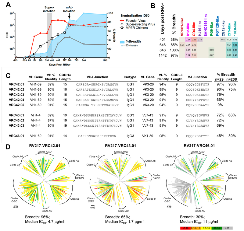
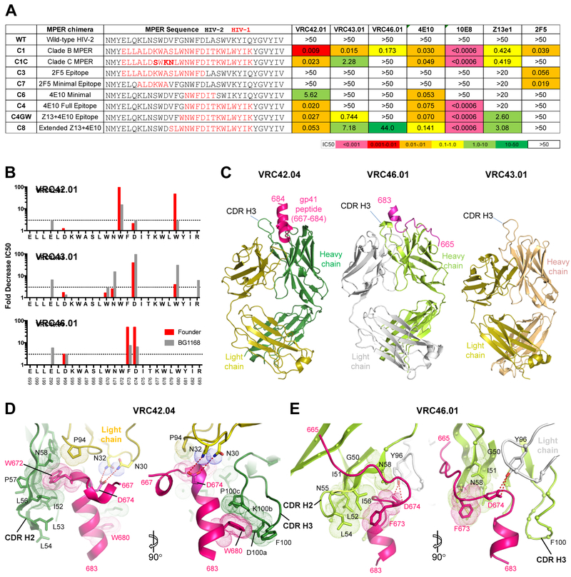
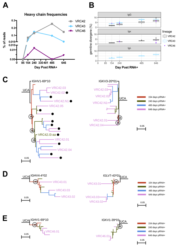
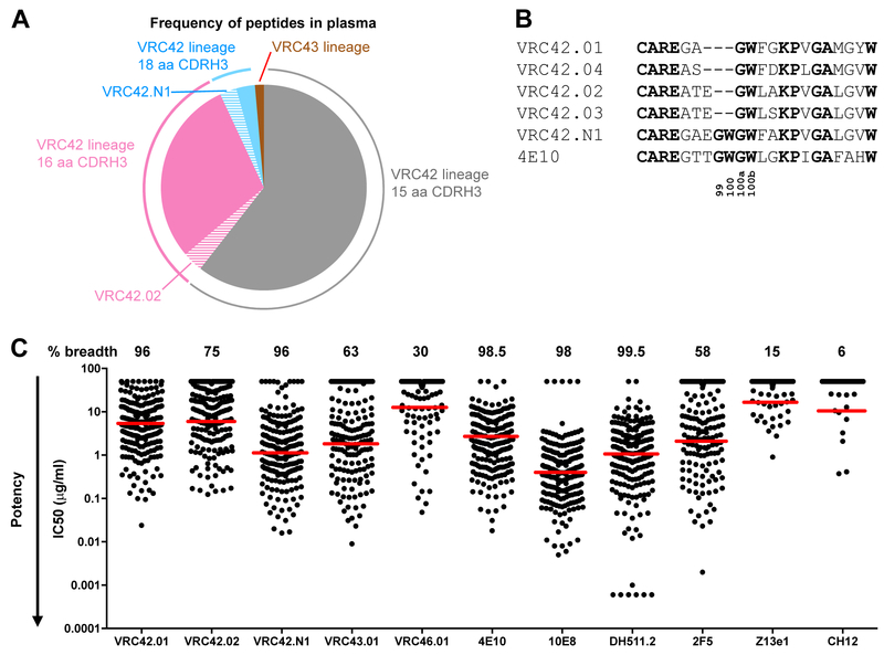
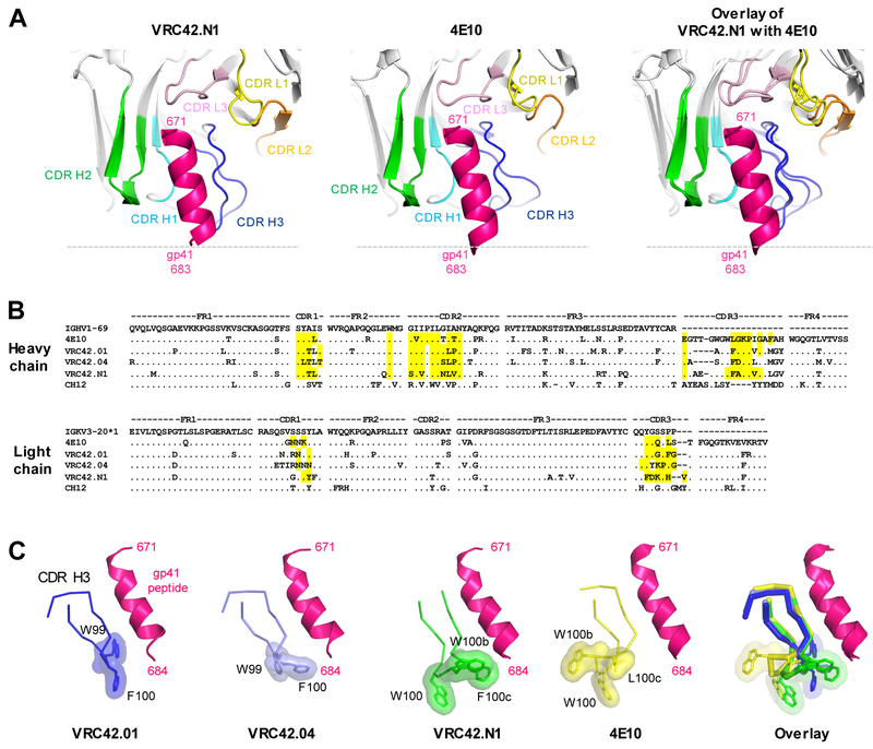
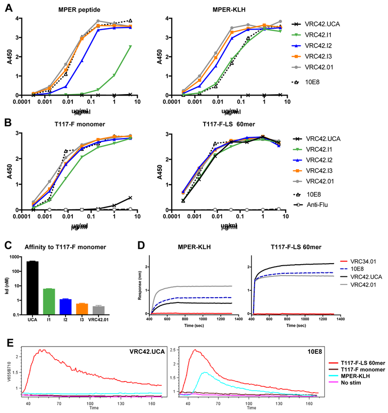
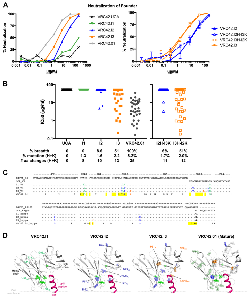
Lineage-based vaccine design is an attractive approach for eliciting broadly neutralizing antibodies (bNAbs) against HIV-1. However, most bNAb lineages studied to date have features indicative of unusual recombination and/or development. From an individual in the prospective RV217 cohort, we identified three lineages of bNAbs targeting the membrane-proximal external region (MPER) of the HIV-1 envelope. Antibodies RV217-VRC42.01, -VRC43.01, and -VRC46.01 used distinct modes of recognition and neutralized 96%, 62%, and 30%, respectively, of a 208-strain virus panel. All three lineages had modest levels of somatic hypermutation and normal antibody-loop lengths and were initiated by the founder virus MPER. The broadest lineage, VRC42, was similar to the known bNAb 4E10. A multimeric immunogen based on the founder MPER activated B cells bearing the unmutated common ancestor of VRC42, with modest maturation of early VRC42 intermediates imparting neutralization breadth. These features suggest that VRC42 may be a promising template for lineage-based vaccine design.

Keywords: HIV envelope; neutralizing antibody; next-generation sequencing.


Figures

Figure 1.
Figure 1.. Donor 40512 from the RV217 cohort achieves broad HIV neutralization through three MPER-directed lineages.
(A) Donor RV217.40512 plasma samples were assessed for neutralization against the founder (red line) and superinfecting (gray line) viruses, HIV-2/HIV1-MPER C1 chimera (dotted line), and heterologous viruses (blue line and fill). Gray bar, window of superinfection. (B) Neutralization-fingerprint analysis using a panel of 34 Env-pseudoviruses. Time point (days) following the first positive RNA test after infection. Values indicate proportion of plasma neutralization associated with the indicated antibody. Colors are defined by epitope (top row) with darker shades indicating higher values. (C) Genetic characterization, isotype, and neutralization breadth of mAbs recovered from B cells at day 646. Percent identity is calculated based on nucleotide sequence. CDR3 length is based on amino acid sequence using Kabat numbering. (D) Neutralization breadth and potency against 208 Env-pseudotyped viruses. Dendrograms show neighbor-joining trees of Env sequences, with branches color-coded by sensitivity to the indicated antibody. See also Table S1 and Figure S1.
Figure 2.
Figure 2.. The Three Lineages Target Distinct Epitopes within the MPER via Different Modes of Recognition.
(A) HIV-2/HIV-1 MPER chimeric viruses were used to determine MPER-specific neutralization. The panel of chimeric viruses is shown with the modified MPER peptide highlighted in red for each chimera (left), and the IC50 values (μg/ml) for each mAb (right). Averages from two experiments are shown. (B) Fold change in IC50 between wild-type founder Env or clade B Env BG1168 viruses containing single ala/gly point mutations. Dotted line, three-fold change. Each assay was performed once. (C) Crystal structures of VRC42.04 Fab and VRC46.01 Fab in complex with the founder MPER peptide, and VRC43.01 Fab alone. (D,E) Close-up views of VRC42.04-MPER (D) and VRC46.01-MPER complex (E). MPER residues that conferred substantial IC50 changes when changed to ala (Figure 2B) are highlighted in magenta. Heavy and light chain residues that are in contact with the highlighted MPER residues were shown in sticks and dots. The Cα atoms of VRC46.01 residues that are in contact with MPER were shown in spheres. The dotted red lines represent hydrogen bonds between two atoms. See also Figures S2-S4 and Tables S3-S5.
Figure 3.
Figure 3.. NGS of B cell Transcripts Reveals All Three Lineages To Be Initiated Between days 85 and 154 Post-Infection but with Divergent Frequencies over Time.
(A) Timing of origin and frequencies of VRC42, VRC43, and VRC46 lineages. % Reads, percent of high-quality reads (see Methods) that are assigned to each lineage. (B) Divergence of heavy and light chains over time from germline precursor V genes. (C) Maximum likelihood phylogenetic trees, based on nucleotide sequence, of the VRC42 lineage showing a representative sampling of the longitudinal NGS data. Colors indicate the time point from which the transcript was sequenced. Dots indicate sequences from which peptides were found in plasma by proteomic analysis at day 646. An NGS sequence with the same amino acid sequence as VRC42.I3-H (VRC42.I3-aa) is labelled in green. Trees for the VRC43 (D) and VRC46 (E) lineages are presented similarly.
Figure 4.
Figure 4.. VRC42 Lineage Member that More Closely Resembles bNAb 4E10 was Identified by Proteomics Analysis.
(A) Proteomic analysis of plasma antibodies detected VRC42 lineage members with CDRH3 lengths of 15 (grey), 16 (pink), and 18 (blue) amino acids at day 646. These included a peptide matching VRC42.02 (16 amino acid CDRH3, pink stripes) and one matching VRC42.N1 (18 amino acid CDRH3, blue stripes). Two peptides from the VRC43 lineage were also detected (brown). Slices are proportional to total peak area, not number of unique peptides. Numbering employs the Kabat convention. (B) Junction and CDRH3 of VRC42.01-.04, NGS-derived sequence VRC42.N1, and 4E10. Residues in bold are identical to 4E10. (C) Neutralization of 208 Env-pseudotyped viruses of diverse clades. Red line, median of neutralized viruses. See also Table S2.
Figure 5.
Figure 5.. VRC42 and 4E10 are Members of the Same bNAb Class.
(A) VRC42.N1 and 4E10 have the same structural mode of recognition. MPER peptide residues (magenta) spanning 671 to 683 of the two structures were superposed and their CDR loops and MPER peptides were displayed in the same angle respect to each other. For clarity, only the MPER region of T117-F in complex with VRC42.N1 is shown. (B) Alignments of heavy and light chain sequences. Top line: predicted naive gene segments for VRC42: VH1-69*10, DH3-10, and JH6. 4E10 and CH12 use VH1-69, but different D and J genes. Residues that contact with gp41 peptide are highlighted in yellow. No structure is available for CH12. (C) Close-up views CDRH3s of VRC42.01, VRC42.04, VRC42.N1, and 4E10 with MPER peptide (magenta). Aromatic residues at the tip were highlighted with surface representation. See also Figure S4 and Tables S4-S5.
Figure 6.
Figure 6.. A Multimeric Immunogen Based on Founder MPER Engages VRC42.UCA and Early Intermediates.
(A-B) Binding of VRC42 lineage antibodies and 10E8 as measured by ELISA. Plates were coated with (A) biotinylated founder MPER peptide or MPER-KLH, (B) T117-F monomer or T117-F-LS 60mer. Data are representative of 2-4 repeat assays. (C) Affinity of VRC42 lineage antibodies to T117-F monomer was measured by Bio-Layer Interferometry (BLI). (D) Binding to MPER-KLH or T117-F-LS 60mer as measured by BLI. (E) Cell signalling through the B cell receptor measured by calcium flux. Cells loaded with the calcium-sensitive dye Fura Red were stimulated with antigen and interrogated by flow cytometry. The ratio of signals in the V655 and B710 channels indicates the level of intracellular calcium-bound Fura Red. Data are representative of 4 repeat assays. See also Figure S5 and S7.
Figure 7.
Figure 7.. VRC42 Intermediates with Low Somatic Mutation have Broad Neutralization Activity.
(A) Neutralization of founder virus by VRC42 lineage intermediates. Data are averages of 6 repeats. (B) Neutralization breadth of VRC42 lineage intermediates to a panel of 29 heterologous pseudoviruses. % mutation is the nucleotide change in heavy and light chains compared to UCA. Data are average of 2 repeat assays. (C) Sequences of intermediates and mature VRC42.01. MPER contact residues are highlighted in yellow. (D) Mapping the somatic mutations on the models (VRC42.I1, I2 and I3) and structure (VRC42.01 mature). Somatic mutations inherited from VRC42.I1, I2, and I3 were shown in green, blue, and orange spheres, respectively. Somatic mutations found in mature VRC42.01 were shown in gray and pale green spheres. See also Figure S6.

Similar articles

Cited by

References

    1. Abbott RK, Lee JH, Menis S, Skog P, Rossi M, Ota T, Kulp DW, Bhullar D, Kalyuzhniy O, Havenar-Daughton C, et al. (2018). Precursor Frequency and Affinity Determine B Cell Competitive Fitness in Germinal Centers, Tested with Germline-Targeting HIV Vaccine Immunogens. Immunity 48, 133–146 e136. - PMC - PubMed
    1. Adams PD, Afonine PV, Bunkoczi G, Chen VB, Davis IW, Echols N, Headd JJ, Hung LW, Kapral GJ, Grosse-Kunstleve RW, et al. (2010). PHENIX: a comprehensive Python-based system for macromolecular structure solution. Acta Crystallogr D Biol Crystallogr 66, 213–221. - PMC - PubMed
    1. Adams PD, Grosse-Kunstleve RW, Hung LW, Ioerger TR, McCoy AJ, Moriarty NW, Read RJ, Sacchettini JC, Sauter NK, and Terwilliger TC (2002). PHENIX: building new software for automated crystallographic structure determination. Acta Crystallogr D Biol Crystallogr 58, 1948–1954. - PubMed
    1. Alam SM, McAdams M, Boren D, Rak M, Scearce RM, Gao F, Camacho ZT, Gewirth D, Kelsoe G, Chen P, et al. (2007). The role of antibody polyspecificity and lipid reactivity in binding of broadly neutralizing anti-HIV-1 envelope human monoclonal antibodies 2F5 and 4E10 to glycoprotein 41 membrane proximal envelope epitopes. J Immunol 178, 4424–4435. - PMC - PubMed
    1. Alam SM, Morelli M, Dennison SM, Liao HX, Zhang R, Xia SM, Rits-Volloch S, Sun L, Harrison SC, Haynes BF, et al. (2009). Role of HIV membrane in neutralization by two broadly neutralizing antibodies. Proc Natl Acad Sci U S A 106, 20234–20239. - PMC - PubMed

Publication types