

Review
doi: 10.1016/j.drudis.2019.03.004.
Epub 2019 Mar 13.
Drug delivery to retinal photoreceptors
Affiliations
- PMID: 30877076
- PMCID: PMC6715772
- DOI: 10.1016/j.drudis.2019.03.004
Item in Clipboard
Review
Drug delivery to retinal photoreceptors
Drug Discov Today.
2019 Aug.
Abstract
The photoreceptors of the retina are afflicted by diseases that still often lack satisfactory treatment options. Although suitable drugs might be available in some cases, the delivery of these compounds into the eye and across the blood-retinal barrier remains a significant challenge for therapy development. Here, we review the routes of drug administration to the retina and highlight different options for drug delivery to the photoreceptor cells.
Copyright © 2019 The Authors. Published by Elsevier Ltd.. All rights reserved.
Figures
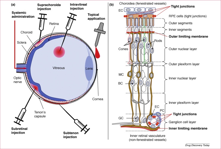
Routes of administration for drugs targeted to the photoreceptors of the retina − illustration of the blood–retinal barrier. (a) Diagrammatic cross-section through an eye, illustrating different routes for administration of drugs to the retina. The ocular cross-section shows on the posterior side (left) the optic nerve, the Tenon capsule surrounding the eye, the sclera, the choroidal vasculature (choroid) and the retina (shown in blue). The anterior side (right) shows the vitreous body, the lens and the cornea. The application routes highlighted are topical, intravitreal, subtenon, suprachoroidal and subretinal injections, as well as systemic administration via the general blood circulation. (b) Idealised cross-section through the retina displaying the choroid and retinal pigment epithelium (RPE; top), the outer and inner nuclear layers, as well as the ganglion cell (GC) layer (bottom). The components of the outer and inner blood–retinal barrier, including the outer and inner limiting membranes, are highlighted in red. Note that the retinal structure has been simplified for clarity and that not all retinal cell types are shown. Abbreviations: BC, bipolar cells; EC, endothelial cell; MC, Müller glial cell; PC, pericyte.
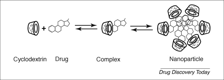
Formation of the solubilising drug–cyclodextrin nanoparticle. The cyclodextrin molecules, which are frequently referred to as host molecules, are displayed as a cup-like structure, which initially forms a complex with individual host molecules. The host shown here represents an idealised dexamethasone molecule. At higher concentrations, drug–cyclodextrin complexes aggregate to form larger nanoparticles that can be administered topically to the eye.
References
-
- Kolb H. How the retina works. Am. Sci. 2003;91:28–35.
-
- Chakravarthy U. The economic impact of blindness in Europe. Ophthal. Epidemiol. 2017;24:239–247. - PubMed
-
- Valdes J. Organotypic retinal explant cultures as in vitro alternative for diabetic retinopathy studies. ALTEX. 2016;33:459–464. - PubMed
-
- McGuinness M.B. Physical activity and age-related macular degeneration: a systematic literature review and meta-analysis. Am. J. Ophthalmol. 2017;180:29–38. - PubMed
-
- Kennan A. Light in retinitis pigmentosa. Trends Genet. 2005;21:103–110. - PubMed
Publication types
MeSH terms
Substances
LinkOut - more resources
Full Text Sources
Medical
