. 2019 Oct;60(10):1444-1451.
doi: 10.2967/jnumed.118.224113. Epub 2019 Mar 15.

Effect of Off-Target Binding on 18F-Flortaucipir Variability in Healthy Controls Across the Life Span

Affiliations

Effect of Off-Target Binding on 18F-Flortaucipir Variability in Healthy Controls Across the Life Span

Suzanne L Baker et al. J Nucl Med. 2019 Oct.

Abstract

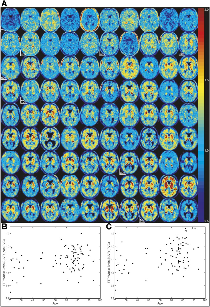
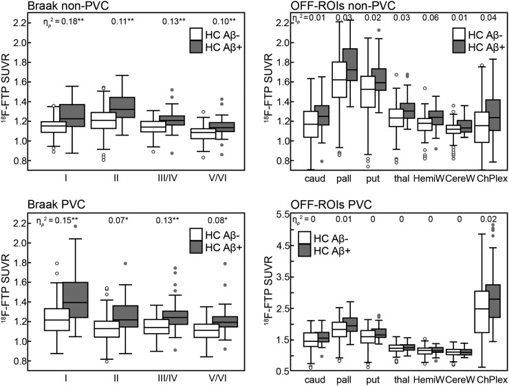
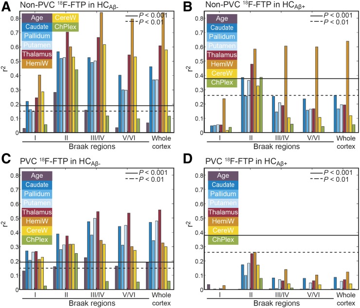
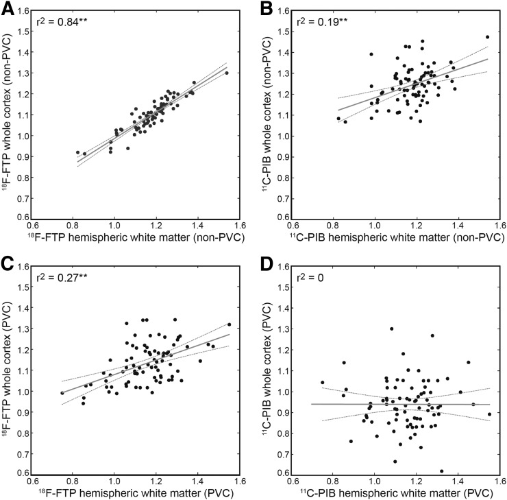
Measuring early tau accumulation is important in studying aging and Alzheimer disease and is only as accurate as the signal-to-noise ratio of the tracer. Along with aggregated tau in the form of neurofibrillary tangles, 18F-flortaucipir has been reported to bind to neuromelanin, monoamine oxidase, calcifications, iron, leptomeningeal melanocytes, and microhemorrages. Although 18F-flortaucipir successfully differentiates healthy controls (HCs) from subjects with Alzheimer disease, variability exists in the cortical signal in amyloid-negative HCs. We aimed to explore the relationship between off-target binding signal and variability in the cortical signal in HCs. Methods: Subjects (n = 139) received 11C-Pittsburgh compound B (PIB) and 18F-flortaucipir PET scans and a magnetization-prepared rapid gradient echo MRI scan. PET frames were realigned and coregistered to the MR images, which were segmented using FreeSurfer. In amyloid-negative HCs (n = 90; age range, 21-94 y), 7 nonspecific or off-target binding regions were considered: caudate, pallidum, putamen, thalamus, cerebellar white matter, hemispheric white matter, and choroid plexus. These regions of interest were assigned to 3 similarly behaving groups using principle components analysis, exploratory factor analysis, and Pearson correlations for caudate, putamen, and pallidum (also correlated with age); thalamus and white matter; and choroid plexus. In amyloid-negative HCs with 11C-PIB and 18F-flortaucipir scans, correlations were calculated between white and gray matter before and after partial-volume correction. Results: The correlation between white and gray matter disappeared after partial-volume correction in 11C-PIB (r2 = 0) but persisted for 18F-flortaucipir (r2 = 0.27), demonstrating that the correlation between white and gray matter signal in 18F-flortaucipir is not solely due to partial-volume effects. A linear regression showed that off-target signal from putamen and thalamus together explained 64% of the variability in the cortical signal in amyloid-negative HCs (not seen in amyloid-positive HCs). Variability in amyloid-negative HCs but not amyloid-positive HCs correlated with white matter signal (unrelated to partial-volume effects) and age-related off-target signal (possibly related to iron load). Conclusion: The noise in the 18F-flortaucipir measurement could pose challenges when studying early tau accumulation.

Keywords: 18F-flortaucipir PET; off-target binding; tau.


Figures

FIGURE 1.
FIGURE 1.
High variability in HCAβ−. (A) 18F-flortaucipir SUVR images in Montreal Neurological Institute space in order of age. Start of each age decade is marked. (B) Age vs. non-PVC whole-cortex SUVR (r2 = 0.06, P < 0.05). (C) Age vs. PVC whole-cortex SUVR (r2 = 0.18, P < 0.001).
FIGURE 2.
FIGURE 2.
Comparison between HCAβ− and HCAβ+ in Braak and OFF ROIs with and without PVC. Central mark indicates median. Top and bottom of box indicate 75th and 25th percentiles, respectively. Whisker extent indicates median ± 1.57(75th percentile – 25th percentile)/square root(number subjects). C represents subjects beyond whisker extent. Significance of P < 0.005 (*) and P < 0.001 (**) denoted for ηp2.
FIGURE 3.
FIGURE 3.
Pearson r2 between 18F-flortaucipir SUVR in cortical regions, age, and OFF ROIs. P values were Bonferroni-corrected.
FIGURE 4.
FIGURE 4.
Correlations between HemiW and whole-cortex SUVRs: non-PVC 18F-flortaucipir (A), non-PVC 11C-PIB (B), PVC 18F-flortaucipir (C), and PVC 11C-PIB (D). Gray line = linear model fit; dashed gray lines = 95% confidence interval. **P < 0.001.

Similar articles

Cited by

  • The Association of Age-Related and Off-Target Retention with Longitudinal Quantification of [18F]MK6240 Tau PET in Target Regions.
    Tissot C, Servaes S, Lussier FZ, Ferrari-Souza JP, Therriault J, Ferreira PCL, Bezgin G, Bellaver B, Leffa DT, Mathotaarachchi SS, Chamoun M, Stevenson J, Rahmouni N, Kang MS, Pallen V, Margherita-Poltronetti N, Wang YT, Fernandez-Arias J, Benedet AL, Zimmer ER, Soucy JP, Tudorascu DL, Cohen AD, Sharp M, Gauthier S, Massarweh G, Lopresti B, Klunk WE, Baker SL, Villemagne VL, Rosa-Neto P, Pascoal TA. Tissot C, et al. J Nucl Med. 2023 Mar;64(3):452-459. doi: 10.2967/jnumed.122.264434. Epub 2022 Nov 17. J Nucl Med. 2023. PMID: 36396455 Free PMC article.
  • Amyloid-PET of the white matter: Relationship to free water, fiber integrity, and cognition in patients with dementia and small vessel disease.
    Ottoy J, Ozzoude M, Zukotynski K, Kang MS, Adamo S, Scott C, Ramirez J, Swardfager W, Lam B, Bhan A, Mojiri P, Kiss A, Strother S, Bocti C, Borrie M, Chertkow H, Frayne R, Hsiung R, Laforce RJ, Noseworthy MD, Prato FS, Sahlas DJ, Smith EE, Kuo PH, Chad JA, Pasternak O, Sossi V, Thiel A, Soucy JP, Tardif JC, Black SE, Goubran M; Medical Imaging Trials Network of Canada (MITNEC) and Alzheimer’s Disease Neuroimaging Initiative (ADNI). Ottoy J, et al. J Cereb Blood Flow Metab. 2023 Jun;43(6):921-936. doi: 10.1177/0271678X231152001. Epub 2023 Jan 25. J Cereb Blood Flow Metab. 2023. PMID: 36695071 Free PMC article.
  • What's the cut-point?: a systematic investigation of tau PET thresholding methods.
    Weigand AJ, Maass A, Eglit GL, Bondi MW. Weigand AJ, et al. Alzheimers Res Ther. 2022 Apr 5;14(1):49. doi: 10.1186/s13195-022-00986-w. Alzheimers Res Ther. 2022. PMID: 35382866 Free PMC article. Review.
  • Translational value of choroid plexus imaging for tracking neuroinflammation in mice and humans.
    Fleischer V, Gonzalez-Escamilla G, Ciolac D, Albrecht P, Küry P, Gruchot J, Dietrich M, Hecker C, Müntefering T, Bock S, Oshaghi M, Radetz A, Cerina M, Krämer J, Wachsmuth L, Faber C, Lassmann H, Ruck T, Meuth SG, Muthuraman M, Groppa S. Fleischer V, et al. Proc Natl Acad Sci U S A. 2021 Sep 7;118(36):e2025000118. doi: 10.1073/pnas.2025000118. Proc Natl Acad Sci U S A. 2021. PMID: 34479997 Free PMC article.
  • Evaluation of [18F]-JNJ-64326067-AAA tau PET tracer in humans.
    Baker SL, Provost K, Thomas W, Whitman AJ, Janabi M, Schmidt ME, Timmers M, Kolb HC, Rabinovici GD, Jagust WJ. Baker SL, et al. J Cereb Blood Flow Metab. 2021 Dec;41(12):3302-3313. doi: 10.1177/0271678X211031035. Epub 2021 Jul 14. J Cereb Blood Flow Metab. 2021. PMID: 34259071 Free PMC article. Clinical Trial.

References

    1. Braak H, Braak E. Neuropathological stageing of Alzheimer-related changes. Acta Neuropathol (Berl). 1991;82:239–259. - PubMed
    1. Chien DT, Bahri S, Szardenings AK, et al. Early clinical PET imaging results with the novel PHF-tau radioligand [F-18]-T807. J Alzheimers Dis. 2013;34:457–468. - PubMed
    1. Xia CF, Arteaga J, Chen G, et al. [18F]T807, a novel tau positron emission tomography imaging agent for Alzheimer’s disease. Alzheimers Dement. 2013;9:666–676. - PubMed
    1. Johnson KA, Schultz A, Betensky RA, et al. Tau positron emission tomographic imaging in aging and early Alzheimer disease. Ann Neurol. 2016;79:110–119. - PMC - PubMed
    1. Schöll M, Lockhart SN, Schonhaut DR, et al. PET imaging of tau deposition in the aging human brain. Neuron. 2016;89:971–982. - PMC - PubMed

Publication types

Substances