. 2019 Jun;68(6):907-915.
doi: 10.1007/s00262-019-02323-x. Epub 2019 Mar 15.

DNA lesions correlate with lymphocyte function after selective internal radiotherapy

Affiliations

DNA lesions correlate with lymphocyte function after selective internal radiotherapy

Aglaia Domouchtsidou et al. Cancer Immunol Immunother. 2019 Jun.

Abstract

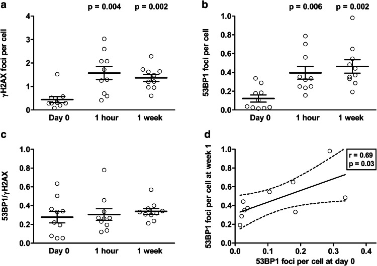
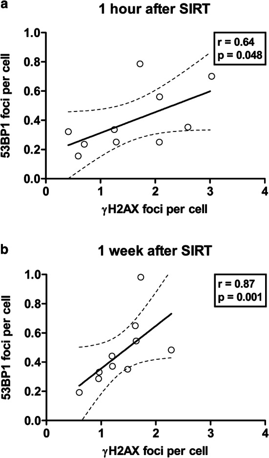
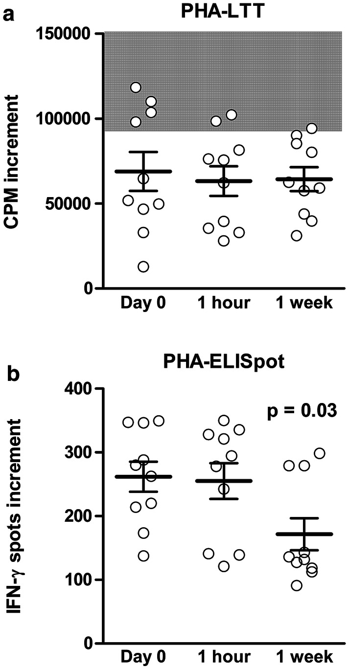
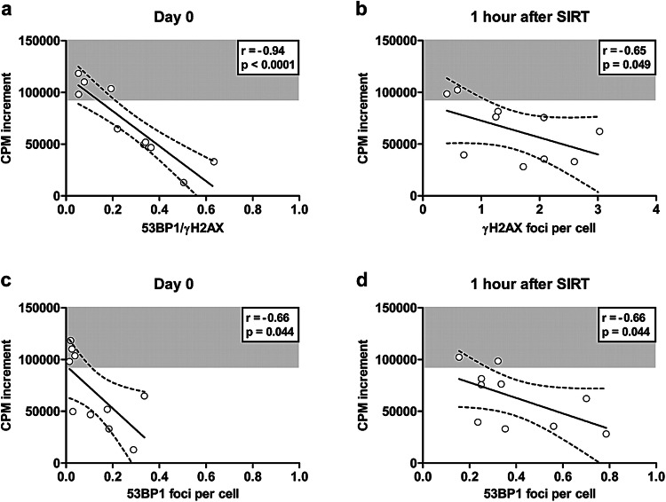
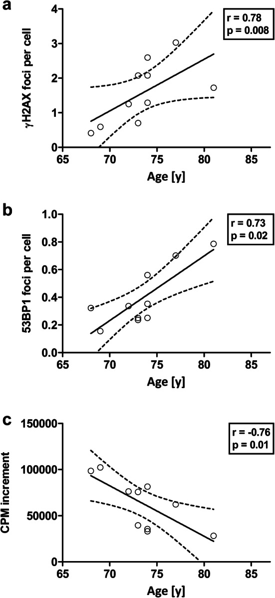
In patients with non-resectable hepatic malignancies selective internal radiotherapy (SIRT) with yttrium-90 is an effective therapy. However, previous data indicate that SIRT leads to impaired immune function. The aim of the current study was to determine the extent of DNA lesions in peripheral blood mononuclear cells of SIRT patients and to correlate these lesions with cellular immune responses. In ten patients γH2AX and 53BP1 foci were determined. These foci are markers of DNA double-strand breaks (DSBs) and occur consecutively. In parallel, lymphocyte proliferation was assessed after stimulation with the T cell mitogen phytohemagglutinin. Analyses of vital cells were performed prior to and 1 h and 1 week after SIRT. 1 h and 1 week after SIRT numbers of γH2AX and of 53BP1 foci were more than threefold larger than before (p < 0.01). Already at baseline, foci were more abundant than published in healthy controls. Lymphocyte proliferation at baseline was below the normal range and further decreased after SIRT. Prior to therapy, there was an inverse correlation between lymphocyte proliferation and the quotient 53BP1/γH2AX; which could be considered as a measure of the course of DNA DSB repair (r = - 0.94, p < 0.0001). Proliferative responses were inversely correlated with 53BP1 foci prior to therapy and γH2AX and 53BP1 foci 1 h after therapy (r < - 0.65, p < 0.05). In conclusion, DNA foci in SIRT patients were correlated with impaired in vitro immune function. Unrepaired DNA DSBs or cell cycle arrest due to repair may cause this impairment.

Keywords: Cellular immune response; DNA double strand break; DNA repair; ELISpot; Lymphocyte proliferation; Selective internal radiotherapy.


Conflict of interest statement

The authors declare that they have no conflict of interest.

Figures

Fig. 1
Fig. 1
DNA foci in ten patients with hepatic malignancies receiving selective internal radiotherapy. γH2AX foci and 53BP1 foci were determined as a measure of DNA double strand breaks. Wilcoxon matched pairs test was used to compare data prior to therapy (day 0) with data 1 h and 1 week after therapy, respectively. Mean and standard error of the mean (SEM) are indicated by horizontal lines (ac). d Shows results of a Spearman correlation analysis between 53BP1 foci at day 0 and week 1 after therapy. The continuous line represents the regression line, the broken lines the 95% confidence interval
Fig. 2
Fig. 2
Spearman correlation analysis of γH2AX and 53BP1 foci in ten patients with hepatic malignancies. Positive correlations were observed a 1 h and b 1 week at after selective internal radiotherapy. The continuous line represents the regression line, the broken lines the 95% confidence interval
Fig. 3
Fig. 3
Lymphocyte responses in ten patients with hepatic malignancies. Lymphocytes were stimulated by the mitogen PHA and a proliferation was determined by lymphocyte transformation test (LTT) and b IFN-γ production by ELISpot. Data are given as counts per minute (CPM) of 3H thymidine uptake or as spot numbers per cell culture. Increment means that the background reaction (without stimulation) was subtracted from the reaction toward PHA. Wilcoxon matched pairs test was used to compare data prior to selective internal radiotherapy (day 0) with data 1 h and 1 week after therapy, respectively. Mean and standard error of the mean (SEM) are indicated by horizontal lines. The gray shaded area displays the normal range which was defined in 177 healthy controls. The threshold is the 5% percentile of cellular responses as determined in these controls
Fig. 4
Fig. 4
Spearman correlation analysis of lymphocyte proliferation and DNA foci in ten patients with hepatic malignancies. Lymphocyte proliferation after stimulation with phythohemagglutinin (PHA) inversely correlates a with the quotient 53BP1/γH2AX prior to selective internal radiotherapy (SIRT) (day 0), b with γH2AX foci 1 h after therapy, c with 53BP1 foci at day 0 and d with 53BP1 foci 1 h after therapy. Data are given as counts per minute (CPM) of 3H thymidine uptake. Increment means that the background reaction (without stimulation) was subtracted from the reaction toward PHA. The continuous line represents the regression line, the broken lines the 95% confidence interval. The gray shaded area displays the normal range which was defined in 177 healthy controls. The threshold is the 5% percentile of cellular responses as determined in these controls
Fig. 5
Fig. 5
Spearman correlation analysis of DNA foci or lymphocyte function and age in ten patients with hepatic malignancies. 1 h after selective internal radiotherapy, there was a significant, positive correlation between a γH2AX foci and age and b 53BP1 foci and age. Furthermore, 1 h after therapy there was an inverse correlation between lymphocyte function and age (c). Lymphocyte function was determined as proliferative response to PHA, given as counts per minute (CPM) of 3H thymidine uptake. Increment means that the background reaction (without stimulation) was subtracted from the reaction toward PHA. The continuous line represents the regression line, the broken lines the 95% confidence interval

References

    1. Lewandowski RJ, Salem R. Yttrium-90 radioembolization of hepatocellular carcinoma and metastatic disease to the liver. Semin Intervent Radiol. 2006;23(1):64–72. doi: 10.1055/s-2006-939842. - DOI - PMC - PubMed
    1. Kennedy AS, McNeillie P, Dezarn WA, et al. Treatment parameters and outcome in 680 treatments of internal radiation with resin 90Y-microspheres for unresectable hepatic tumors. Int J Radiat Oncol Biol Phys. 2009;74(5):1494–1500. doi: 10.1016/j.ijrobp.2008.10.005. - DOI - PubMed
    1. Wang LM, Jani AR, Hill EJ, Sharma RA. Anatomical basis and histopathological changes resulting from selective internal radiotherapy for liver metastases. J Clin Pathol. 2013;66(3):205–211. doi: 10.1136/jclinpath-2012-201231. - DOI - PMC - PubMed
    1. Dezarn WA, Cessna JT, DeWerd LA, et al. Recommendations of the American Association of Physicists in Medicine on dosimetry, imaging, and quality assurance procedures for 90Y microsphere brachytherapy in the treatment of hepatic malignancies. Med Phys. 2011;38(8):4824–4845. doi: 10.1118/1.3608909. - DOI - PubMed
    1. Salem R, Thurston KG. Radioembolization with 90yttrium microspheres: a state-of-the-art brachytherapy treatment for primary and secondary liver malignancies. Part 2: special topics. J Vasc Interv Radiol. 2006;17(9):1425–1439. doi: 10.1097/01.RVI.0000235779.88652.53. - DOI - PubMed

MeSH terms

LinkOut - more resources