. 2019 May;138(5):525-533.
doi: 10.1007/s00439-019-01997-8. Epub 2019 Mar 15.

Canine models of human amelogenesis imperfecta: identification of novel recessive ENAM and ACP4 variants

Affiliations

Canine models of human amelogenesis imperfecta: identification of novel recessive ENAM and ACP4 variants

Marjo K Hytönen et al. Hum Genet. 2019 May.

Abstract

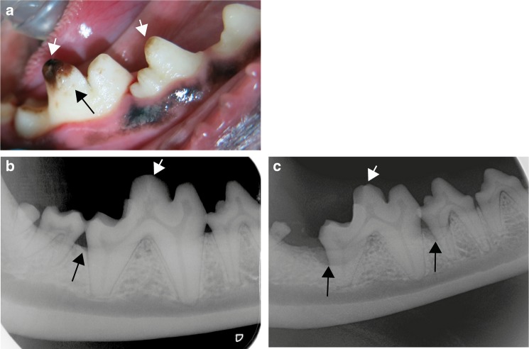
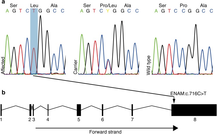
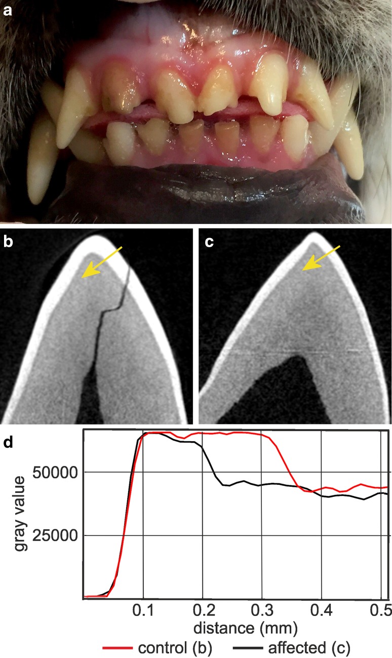
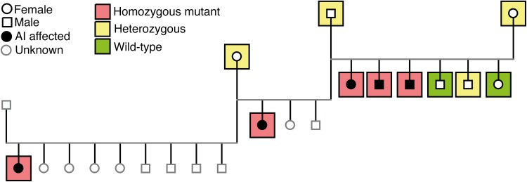
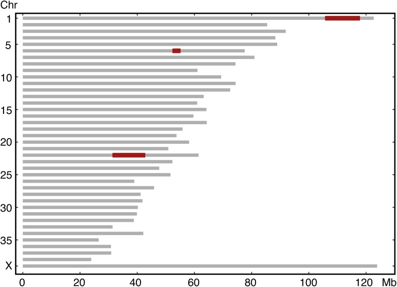
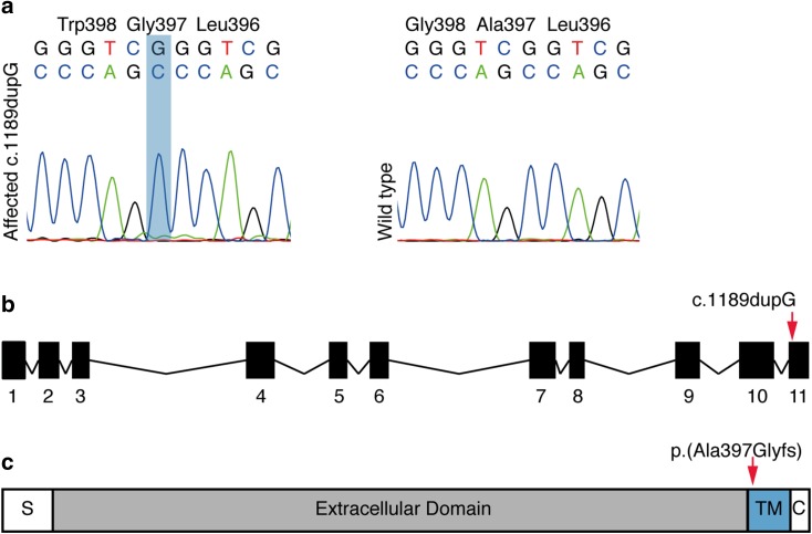
Amelogenesis imperfecta (AI) refers to a genetically and clinically heterogeneous group of inherited disorders affecting the structure, composition, and quantity of tooth enamel. Both non-syndromic and syndromic forms of AI have been described and several genes affecting various aspects of the enamel physiology have been reported. Genetically modified murine models of various genes have provided insights into the complex regulation of proper amelogenesis. Non-syndromic AI occurs spontaneously also in dogs with known recessive variants in ENAM and SLC24A4 genes. Unlike rodents with a reduced dentition and continuously erupting incisors, canine models are valuable for human AI due to similarity in the dental anatomy including deciduous and permanent teeth. We have performed a series of clinical and genetic analyses to investigate AI in several breeds of dogs and describe here two novel recessive variants in the ENAM and ACP4 genes. A fully segregating missense variant (c.716C>T) in exon 8 of ENAM substitutes a well-conserved proline to leucine, p.(Pro239Leu), resulting in a clinical hypomineralization of teeth. A 1-bp insertion in ACP4 (c.1189dupG) is predicted to lead to a frameshift, p.(Ala397Glyfs), resulting in an abnormal C-terminal part of the protein, and hypoplastic AI. The ENAM variant was specific for Parson Russell Terriers with a carrier frequency of 9%. The ACP4 variant was found in two breeds, Akita and American Akita with a carrier frequency of 22%. These genetic findings establish novel canine models of human AI with a particular interest in the case of the ACP4-deficient model, since ACP4 physiology is poorly characterized in human AI. The affected dogs could also serve as preclinical models for novel treatments while the breeds would benefit from genetic tests devised here for veterinary diagnostics and breeding programs.


Conflict of interest statement

HL has consulted Genoscoper Laboratories Oy, which provides genetic tests for dogs, including the findings in this study.

Figures

Fig. 1
Fig. 1
Clinical features of amelogenesis imperfecta in Parson Russell Terriers. a A photograph of the right mandibular PM4 and M1 tooth of Dog 3 at 4 years of age after the teeth had been scaled and polished. The enamel is uneven and spotted with tiny pits (black arrow). At the crown tip there is abrasion and staining of the exposed dentin (white arrows). b A dental radiograph of the right mandibular PM4, M1 and M2 teeth of Dog 3 at 4 years of age. There is very mild alveolar bone loss distally to M1 (black arrow). The cusp tip of M1 is flattened from abrasion (white arrow). c A dental radiograph of the right mandibular PM4 and M1 teeth of Dog 3 at age of 6.5 years. There is moderate alveolar bone loss at the distal root of PM4 and M1 (black arrows). The cusp tip of M1 is flattened from abrasion (long arrow). M2 has been earlier extracted due to periodontitis
Fig. 2
Fig. 2
Identification of a missense variant in ENAM associated with AI in PRTs. a The Sanger sequencing chromatograms showing ENAM:c.716C>T variant as homozygous in affected dog and heterozygous and wild-type in carrier and normal, respectively. bENAM gene contains eight exons and the variant c.716C>T is located in the exon 8
Fig. 3
Fig. 3
Enamel hypoplasia in Akitas. a A photograph illustrating hypoplastic AI with severe abrasion of incisors in a 7-month old Akita. The photograph used with permission from Eeva Länsisola. b MicroCT analysis of a mandibular right deciduous third premolar (DP3) of a healthy Akita. Posterior view of the major cusp. c MicroCT analysis of a mandibular right deciduous third premolar (DP3) of an affected Akita. Posterior view of the major cusp. The enamel layer is generally thinner than in the healthy tooth. d Plots of mineral density of the control and affected teeth across the lines indicated by the yellow arrows in b, c. On the labial side of the cusp tip the enamel thickness of the affected tooth is reduced to 50% of control. Mineralization of the enamel is slightly reduced while the dentin appears normal
Fig. 4
Fig. 4
Pedigree constructed around five Akitas affected by amelogenesis imperfecta. The affected dogs are closely related and both females and males are affected. The ACP4 variant segregated in the pedigree according to a recessively inherited disease
Fig. 5
Fig. 5
Homozygosity mapping resulted in three ROHs, in chr1 (105,875,051–117,668,302 bp), 6 (52,522,808–54,942,283 bp) and 22 (31,559,668–42,601,865 bp) (marked in red)
Fig. 6
Fig. 6
Identification of a duplication in ACP4 associated in AI in Akitas. The Sanger sequencing chromatograms showing c.1189dupG variant as homozygous in the affected dog and wild-type in the unaffected dog. bACP4 gene contains 11 exons and the variant is located in the last exon. c The variant is predicted to lead to a frameshift changing the amino acid sequence starting from the extracellular end of the transmembrane domain (amino acids 395–415)

References

    1. Adzhubei IA, Schmidt S, Peshkin L, Ramensky VE, Gerasimova A, Bork P, Kondrashov AS, Sunyaev SR. A method and server for predicting damaging missense mutations. Nat Methods. 2010;7:248–249. doi: 10.1038/nmeth0410-248. - DOI - PMC - PubMed
    1. Aldred MJ, Savarirayan R, Crawford PJ. Amelogenesis imperfecta: a classification and catalogue for the 21st century. Oral Dis. 2003;9:19–23. doi: 10.1034/j.1601-0825.2003.00843.x. - DOI - PubMed
    1. Backman B, Holm AK. Amelogenesis imperfecta: prevalence and incidence in a northern Swedish county. Community Dent Oral Epidemiol. 1986;14:43–47. doi: 10.1111/j.1600-0528.1986.tb01493.x. - DOI - PubMed
    1. Broeckx BJ, Coopman F, Verhoeven GE, Bavegems V, De Keulenaer S, De Meester E, Van Niewerburgh F, Deforce D. Development and performance of a targeted whole exome sequencing enrichment kit for the dog (Canis Familiaris Build 3.1) Sci Rep. 2014;4:5597. doi: 10.1038/srep05597. - DOI - PMC - PubMed
    1. Broeckx BJ, Hitte C, Coopman F, Verhoeven GE, De Keulenaer S, De Meester E, Derrien T, Alfoldi J, Lindblad-Toh K, Bosmans T, Gielen I, Van Bree H, Van Ryssen B, Saunders JH, Van Nieuwerburgh F, Deforce D. Improved canine exome designs, featuring ncRNAs and increased coverage of protein coding genes. Sci Rep. 2015;5:12810. doi: 10.1038/srep12810. - DOI - PMC - PubMed

LinkOut - more resources