The Role of Redox Dysregulation in the Effects of Prenatal Stress on Embryonic Interneuron Migration
- PMID: 30877797
- PMCID: PMC7199998
- DOI: 10.1093/cercor/bhz052
The Role of Redox Dysregulation in the Effects of Prenatal Stress on Embryonic Interneuron Migration
Abstract
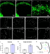
Maternal stress during pregnancy is associated with increased risk of psychiatric disorders in offspring, but embryonic brain mechanisms disrupted by prenatal stress are not fully understood. Our lab has shown that prenatal stress delays inhibitory neural progenitor migration. Here, we investigated redox dysregulation as a mechanism for embryonic cortical interneuron migration delay, utilizing direct manipulation of pro- and antioxidants and a mouse model of maternal repetitive restraint stress starting on embryonic day 12. Time-lapse, live-imaging of migrating GAD67GFP+ interneurons showed that normal tangential migration of inhibitory progenitor cells was disrupted by the pro-oxidant, hydrogen peroxide. Interneuron migration was also delayed by in utero intracerebroventricular rotenone. Prenatal stress altered glutathione levels and induced changes in activity of antioxidant enzymes and expression of redox-related genes in the embryonic forebrain. Assessment of dihydroethidium (DHE) fluorescence after prenatal stress in ganglionic eminence (GE), the source of migrating interneurons, showed increased levels of DHE oxidation. Maternal antioxidants (N-acetylcysteine and astaxanthin) normalized DHE oxidation levels in GE and ameliorated the migration delay caused by prenatal stress. Through convergent redox manipula-tions, delayed interneuron migration after prenatal stress was found to critically involve redox dysregulation. Redox biology during prenatal periods may be a target for protecting brain development.
Keywords: N-acetylcysteine; antioxidants; interneuron migration; oxidative stress; prenatal stress.
© The Author(s) 2019. Published by Oxford University Press. All rights reserved. For Permissions, please e-mail: journals.permissions@oup.com.
Figures


Similar articles
-
The Impact of Maternal Antioxidants on Prenatal Stress Effects on Offspring Neurobiology and Behavior.Yale J Biol Med. 2022 Mar 31;95(1):87-104. eCollection 2022 Mar. Yale J Biol Med. 2022. PMID: 35370489 Free PMC article.
-
Persistent Interneuronopathy in the Prefrontal Cortex of Young Adult Offspring Exposed to Ethanol In Utero.J Neurosci. 2015 Aug 5;35(31):10977-88. doi: 10.1523/JNEUROSCI.1462-15.2015. J Neurosci. 2015. PMID: 26245961 Free PMC article.
-
Delays in GABAergic interneuron development and behavioral inhibition after prenatal stress.Dev Neurobiol. 2016 Oct;76(10):1078-91. doi: 10.1002/dneu.22376. Epub 2016 Jan 22. Dev Neurobiol. 2016. PMID: 26724783 Free PMC article.
-
An Overview of the Mechanisms of Abnormal GABAergic Interneuronal Cortical Migration Associated with Prenatal Ethanol Exposure.Neurochem Res. 2017 May;42(5):1279-1287. doi: 10.1007/s11064-016-2169-5. Epub 2017 Feb 3. Neurochem Res. 2017. PMID: 28160199 Review.
-
Cellular stress mechanisms of prenatal maternal stress: Heat shock factors and oxidative stress.Neurosci Lett. 2019 Sep 14;709:134368. doi: 10.1016/j.neulet.2019.134368. Epub 2019 Jul 9. Neurosci Lett. 2019. PMID: 31299286 Free PMC article. Review.
Cited by
-
Caught in vicious circles: a perspective on dynamic feed-forward loops driving oxidative stress in schizophrenia.Mol Psychiatry. 2022 Apr;27(4):1886-1897. doi: 10.1038/s41380-021-01374-w. Epub 2021 Nov 10. Mol Psychiatry. 2022. PMID: 34759358 Free PMC article. Review.
-
Maternal P7C3-A20 Treatment Protects Offspring from Neuropsychiatric Sequelae of Prenatal Stress.Antioxid Redox Signal. 2021 Sep 1;35(7):511-530. doi: 10.1089/ars.2020.8227. Epub 2021 Jan 29. Antioxid Redox Signal. 2021. PMID: 33501899 Free PMC article.
-
Environmental Enrichment Promotes Transgenerational Programming of Uterine Inflammatory and Stress Markers Comparable to Gestational Chronic Variable Stress.Int J Mol Sci. 2023 Feb 13;24(4):3734. doi: 10.3390/ijms24043734. Int J Mol Sci. 2023. PMID: 36835144 Free PMC article.
-
Threshold effects of prenatal stress on striatal microglia and relevant behaviors.bioRxiv [Preprint]. 2025 Jan 30:2025.01.30.635666. doi: 10.1101/2025.01.30.635666. bioRxiv. 2025. PMID: 39975105 Free PMC article. Preprint.
-
Prenatal stress alters mouse offspring dorsal striatal development and placental function in sex-specific ways.J Psychiatr Res. 2025 Feb;182:149-160. doi: 10.1016/j.jpsychires.2024.12.048. Epub 2025 Jan 1. J Psychiatr Res. 2025. PMID: 39809011
References
-
- Al-Amin MM, Alam T, Hasan SM, Hasan AT, Quddus AH. 2016. Prenatal maternal lipopolysaccharide administration leads to age- and region-specific oxidative stress in the early developmental stage in offspring. Neuroscience. 318:84–93. - PubMed
-
- Al-Amin MM, Reza HM, Saadi HM, Mahmud W, Ibrahim AA, Alam MM, Kabir N, Saifullah AR, Tropa ST, Quddus AH. 2016. Astaxanthin ameliorates aluminum chloride-induced spatial memory impairment and neuronal oxidative stress in mice. Eur J Pharmacol. 777:60–69. - PubMed
-
- Azuelos I, Jung B, Picard M, Liang F, Li T, Lemaire C, Giordano C, Hussain S, Petrof BJ. 2015. Relationship between autophagy and ventilator-induced diaphragmatic dysfunction. Anesthesiology. 122:1349–1361. - PubMed
Publication types
MeSH terms
Substances
Grants and funding
LinkOut - more resources
Full Text Sources
Medical
Miscellaneous

