. 2019 May;95(5):1167-1180.
doi: 10.1016/j.kint.2018.11.043. Epub 2019 Mar 8.

Matrix metalloproteinase-7 protects against acute kidney injury by priming renal tubules for survival and regeneration

Affiliations

Matrix metalloproteinase-7 protects against acute kidney injury by priming renal tubules for survival and regeneration

Haiyan Fu et al. Kidney Int. 2019 May.

Abstract

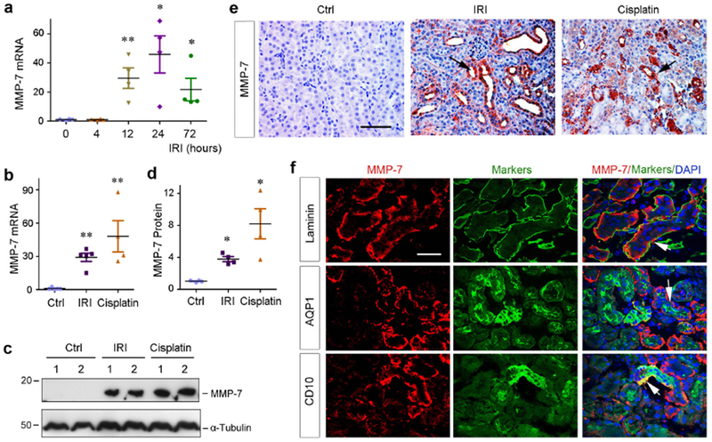
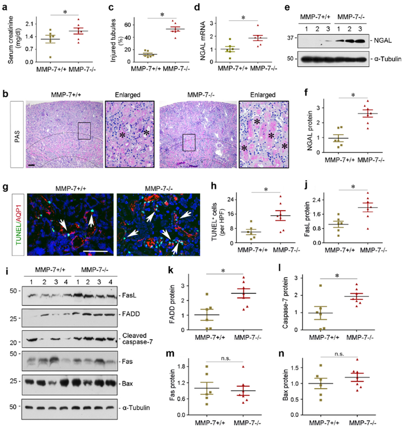
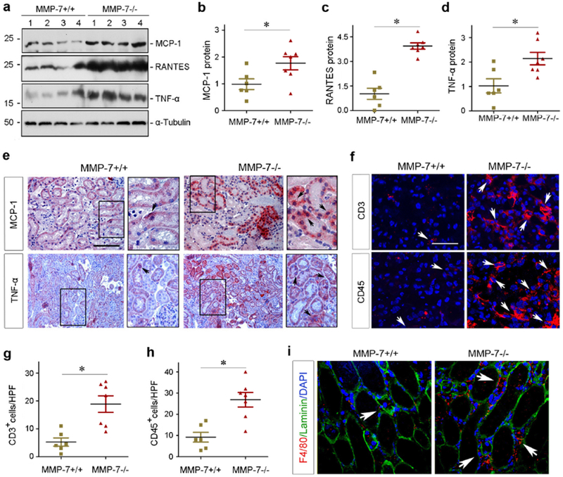
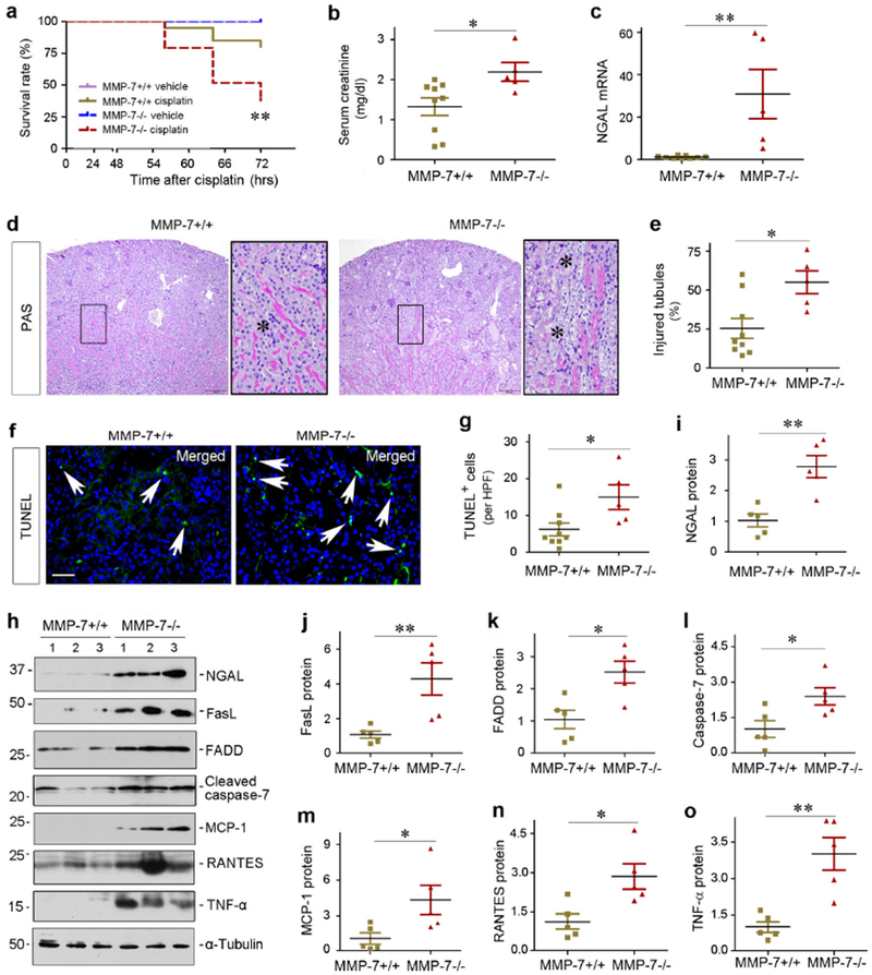
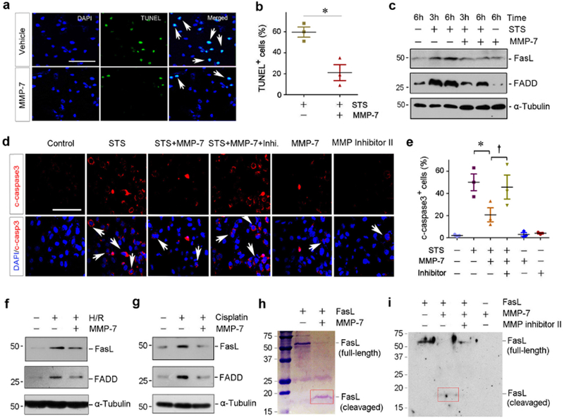
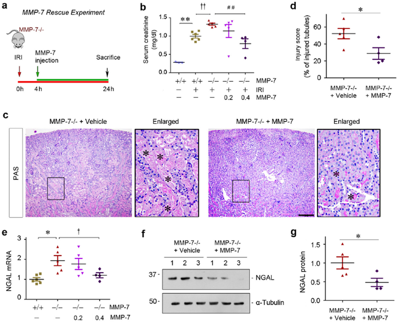
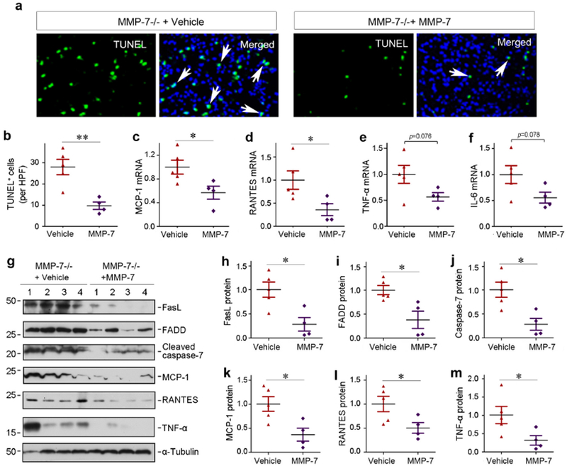
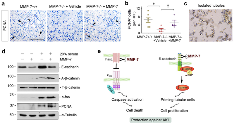
Matrix metalloproteinase-7 (MMP-7) is a secreted endopeptidase that degrades a broad range of substrates. Recent studies have identified MMP-7 as an early biomarker to predict severe acute kidney injury (AKI) and poor outcomes after cardiac surgery; however, the role of MMP-7 in the pathogenesis of AKI is unknown. In this study, we investigated the expression of MMP-7 and the impact of MMP-7 deficiency in several models of AKI. MMP-7 was induced in renal tubules following ischemia/ reperfusion injury or cisplatin administration, and in folic acid-induced AKI. MMP-7 knockout mice experienced higher mortality, elevated serum creatinine, and more severe histologic lesions after ischemic or toxic insults. Tubular apoptosis and interstitial inflammation were more prominent in MMP-7 knockout kidneys. These histologic changes were accompanied by increased expression of FasL and other components of the extrinsic apoptotic pathway, as well as increased expression of pro-inflammatory chemokines. In a rescue experiment, exogenous MMP-7 ameliorated kidney injury in MMP-7 knockout mice after ischemia/reperfusion. In vitro, MMP-7 protected tubular epithelial cells against apoptosis by directly degrading FasL. In isolated tubules ex vivo, MMP-7 promoted cell proliferation by degrading E-cadherin and thereby liberating β-catenin, priming renal tubules for regeneration. Taken together, these results suggest that induction of MMP-7 is protective in AKI by degrading FasL and mobilizing β-catenin, thereby priming kidney tubules for survival and regeneration.

Keywords: FasL; MMP-7; acute kidney injury; apoptosis; proliferation; β-catenin.


Figures

Figure 1.
Figure 1.
Induction of MMP-7 is a common finding in various AKI. (a) Time-dependent induction of MMP-7 in mouse kidneys after IRI. Total RNA was isolated from control kidney and injured kidneys at different time points after IRI as indicated. Relative abundance of renal MMP-7 mRNA was assessed by qRT-PCR, and fold induction over the controls was reported. **P<0.01, *P<0.05 versus sham controls (n=4). (b) Both ischemic and toxic AKI induces renal MMP-7 mRNA expression. Relative level of renal MMP-7 mRNA was assessed by qRT-PCR at different time points after IRI (1 day) and cisplatin (3 days), respectively. **P< 0.01 vs sham controls (n=4-5). (c, d) Western blot analyses of renal expression of MMP-7 protein in injured kidneys after AKI induced by IRI and cisplatin. Representative western blot (c) and quantitative data (d) were presented. Numbers (1-2) indicate each individual animal in a given group. *P< 0.05 vs sham controls. (e) Representative micrographs show MMP-7 protein expression and localization in control and injured kidneys at 1 day after IRI or 3 days after cisplatin. Arrows indicate positive staining. Scale bar, 60 μm. (f) Co-immunostaining shows tubular localization of MMP-7 after IRI. Cryosections from the kidney at 1 day after IRI were double-stained with antibodies against MMP-7 (red) or different markers (green) including laminin, AQP1 and CD10, respectively. Arrows indicate positive staining. Scale bar, 50 μm.
Figure 2.
Figure 2.
Deletion of endogenous MMP-7 aggravates ischemic AKI in mice. (a) Serum creatinine level in MMP-7+/+ wild-type and MMP-7−/− null mice at 1 day after IRI. *P< 0.05 (n=6-7). (b) Representative micrographs show kidney morphology in MMP-7+/+ and MMP-7−/− mice at 1 day after IRI. Asterisks in the enlarged boxed areas indicate injured tubules. Dashed lines show renal corticomedullary junction area. Scale bar, 50 μm. (c) Semi-quantitative assessment of kidney injury (percentage of injured tubules) at 1 day after IRI. *P< 0.05 (n=6-7). (d) qRT-PCR analyses show an increased NGAL mRNA expression in the injured kidneys of MMP-7−/− mice at 1 day after IRI, compared with MMP-7+/+ controls. *P< 0.05 (n=6-7). (e, f) Western blot analyses of renal NGAL protein in MMP-7+/+ and MMP-7−/− mice at 1 day after IRI. Representative western blot (e) and quantitative data (f) are presented. *P < 0.05 (n=6-7). Numbers (1-3) indicate each individual animal in a given group. (g) Representative micrographs show apoptotic proximal tubular cells detected by co-staining of TUNEL (green) and AQP1 (red) in MMP-7+/+ and MMP-7−/− mice. Arrows indicate apoptotic cells. Scale bar, 100 pm. (h) Quantitative determination of apoptotic cells at 1 day after IRI. Data are presented as numbers of apoptotic cells per high power field (HPF). *P< 0.05 (n=6-7). (i) Representative western blotting show the protein levels of renal FasL, FADD, cleaved caspase-7, Fas and Bax. Numbers (1-4) indicate each individual animal in a given group. (j-n) Quantitative determination of renal protein levels of FasL (j), FADD (k), cleaved caspase-7 (l), Fas (m) and Bax (n) in MMP-7+/+ and MMP-7−/− mice at 1 days after IRI. *P< 0.05 (n=6-7).
Figure 3.
Figure 3.
Ablation of endogenous MMP-7 induces pro-inflammatory cytokine expression and promotes renal inflammation after ischemic AKI. (a-d) Western blot analyses of MCP-1, RANTES, and TNF-α protein abundance in MMP-7+/+ and MMP-7−/− kidneys at 1 day after IRI. Representative western blot (a) and quantitative data (b-d) were presented. Numbers (1-4) indicated each individual animal in a given group. *P < 0.05 (n=6-7). (e) Representative micrographs show MCP-1 and TNF-α protein in MMP-7+/+ and MMP-7−/− kidneys at 1 day after IRI. Arrows in the enlarged boxes indicate positive staining. Scale bar, 100 μm. (f) Immunofluorescence staining reveals an increased infiltration of CD3+ T cells and CD45+ leukocytes in MMP-7−/− kidneys at 1 day after IRI. Arrows indicate positive staining. Scale bar, 50 μm. (g, h) Quantitative data show the numbers of CD3+ (g) or CD45+ cells (h) per high-power field (HPF). *P< 0.05 (n=6-7). (i) Co-immunofluorescence staining for F4/80 (red), basement membrane protein laminin (green) and nuclei (blue) shows an increased macrophages infiltration at 1 day after IRI in MMP-7−/− mice, compared with MMP-7+/+ controls. Arrows indicate interstitial macrophages. Scale bar, 50 μm.
Figure 4.
Figure 4.
Loss of endogenous MMP-7 aggravates toxic AKI induced by cisplatin. (a) Survival curve shows higher mortality rate in MMP-7−/− mice at 3 days after cisplatin, compared with MMP-7+/+ controls. **P< 0.01 (n=20-29). (b) Serum creatinine level in MMP-7+/+ and MMP-7−/− mice at 3 days after cisplatin injection. *P< 0.05 (n=5-9). (c) qRT-PCR analysis reveals an increased NGAL mRNA in injured kidneys of MMP-7−/− mice at 3 days after cisplatin, compared with MMP-7+/+ controls. **P< 0.01 (n=5-9). **P< 0.01 (n=5-9). (d) Representative micrographs of the kidneys in MMP-7+/+ and MMP-7−/− mice at 3 days after cisplatin. Asterisks in the enlarged boxed areas indicate injured tubules. Scale bar, 150 μm. (e) Injured tubules in MMP-7+/+ and MMP-7−/− mice at 3 days after cisplatin. The percentages of injured tubules (%) are presented. *P< 0.05 (n=5-9). (f) Representative micrographs show apoptotic cells detected by staining in MMP-7+/+ and MMP-7−/− mice at 3 days after cisplatin. Arrows indicate apoptotic cells. Scale bar, 50 μm. (g) Quantitative determination of apoptotic cells at 3 days after cisplatin. Data are presented as numbers of apoptotic cells per high power field (HPF). *P< 0.05 (n=5-9). (h) Representative western blot analyses of various proteins in the injured kidneys of MMP-7+/+ and MMP-7−/− mice at 3 days after cisplatin. Numbers (1-3) indicate each individual animal in a given group. (i-o) Quantitative determination of the relative protein levels in MMP-7+/+ and MMP-7−/− kidneys at 3 days after cisplatin. Proteins of NGAL (i), FasL (j), FADD (k), caspase-7 (l), MCP-1 (m), RANTES (n) and TNF-α (o) are presented. **P< 0.01, *P< 0.05 (n=5).
Figure 5.
Figure 5.
MMP-7 protects kidney tubular cells against apoptosis by cleaving FasL in vitro. (a) Human recombinant MMP-7 protein protects tubular cells against apoptosis induced by staurosporine (STS). HKC-8 cells were incubated with recombinant MMP-7 or vehicle, followed by treatment with STS (0.1 nM) as indicated. Apoptosis was assessed by TUNEL staining. Representative micrographs of TUNEL staining (a) and quantitative data of apoptotic cells are presented (b). Arrows indicate apoptotic cells. *P< 0.05 vs vehicle (n=3). (c) Representative western blotting shows that MMP-7 decreased FasL and FADD protein in HKC-8 cells following STS treatment. (d, e) MMP-7 modulates caspase-3 activation in HKC-8 cells after STS, which is dependent on its enzymatic activity. HKC-8 cells were treated with STS in the absence or presence of MMP-7 and MMP inhibitor II as indicated. Representative micrographs of immunostaining for cleaved caspase-3 (d) and quantitative data (e) are presented. *P< 0.05 vs vehicle (n=3). P< 0.05 vs MMP-7 without MMP inhibitor II (n=3). Scale bar, 100 μm. Arrows indicate positive staining. (f) Representative western blotting shows that MMP-7 abolished FasL and FADD expression induced by hypoxia/reoxygenation (H/R). HKC-8 cells were incubated in the absence or presence of MMP-7 protein (25 nM) in hypoxic condition for 6 hours, followed by reoxygenation for 2 hours. (g) Representative western blotting shows that MMP-7 abolished FasL and FADD protein expression induced by cisplatin. HKC-8 cells were incubated with cisplatin (25 μg/ml) for 24 hours in the absence or presence of MMP-7 protein (25 nM). (h) Representative SDS-PAGE shows that MMP-7 directly degrades FasL. Recombinant mouse FasL (2 pg) was incubated with 50 nM MMP-7 for 60 min. Full-length FasL migrates at the location of ~50 kDa. Red box indicates the degraded fragments of FasL. (i) Western blot analyses show FasL and its degraded fragments after incubation with MMP-7 in the absence or presence of MMP inhibitor II. FasL degraded fragment is indicated by Red box.
Figure 6.
Figure 6.
Exogenous MMP-7 rescues its renoprotection after AKI in MMP-7−/− null mice. (a) Rescue experiment design. At 4 h after IRI, a single dose of MMP-7 was injected intravenously, and mice were sacrificed at 24 h. (b) Serum creatinine level in MMP-7+/+ and MMP-7−/− mice injected with either vehicle or recombinant MMP-7 at different dosage after IRI. **P< 0.01 vs sham control (n=3-6); ††P< 0.01 vs MMP-7+/+ mice (n=5-6); ##P< 0.01 vs MMP-7−/− mice (n=4-5) with exogenous MMP-7. (c) Representative micrographs show morphological injury of the kidneys in MMP-7−/− mice injected with vehicle or MMP-7. Asterisks in the enlarged boxed areas indicate injured tubules. Scale bar, 200 μm. (d) Quantitative assessment of morphological injury is presented. *P< 0.05 (n=4-5). (e) qRT-PCR analyses show renal NGAL mRNA expression in various groups as indicated. *P< 0.05 vs sham control; P<0.05 vs MMP-7−/− mice with exogenous MMP-7 (n=4-6). (f, g) Western blot analyses of renal NGAL protein in MMP-7−/− mice injected with vehicle or MMP-7. Representative western blot (f) and quantitative data (g) were presented. Numbers (1-3) indicate each individual animal in a given group. *P< 0.05 (n=4-5).
Figure 7.
Figure 7.
Exogenous MMP-7 ameliorates apoptosis and renal inflammation in MMP-7−/− null mice in vivo. (a, b) TUNEL staining shows a reduced apoptosis in the injured kidneys of MMP-7−/− mice injected exogenous MMP-7 after IRI. Representative micrographs (a) and quantitative data (b) are presented. Arrows in the TUNEL/DAPI merged images indicate apoptotic tubular cells. **P< 0.01 (n=4-5). (c-f) qRT-PCR analyses show the relative mRNA levels of renal MCP-1, RANTES, TNF-α, and IL-6 in MMP-7−/− mice injected with vehicle or exogenous MMP-7 after IRI. *P< 0.05 (n=4-5). (g) Representative western blotting show a reduced expression of both apoptosis-regulatory proteins and pro-inflammatory chemokines in MMP-7−/− mice injected with vehicle or exogenous MMP-7. (h-m) Quantitative determination of the relative protein levels of renal FasL, FADD, cleaved caspase-7, MCP-1, RANTES and TNF-α. Numbers (1-4) indicated each individual animal in a given group. *P < 0.05 (n=4-5).
Figure 8.
Figure 8.
MMP-7 promotes tubular cell proliferation after AKI in vivo and ex vivo. (a) Representative micrographs show PCNA+ tubular cells in MMP-7−/− mice injected with vehicle or exogenous MMP-7 after IRI. (b) Quantitative determination of PCNA+ tubular cells per high power field (HPF) at 1 day after IRI. *P < 0.05 vs MMP-7+/+ mice (n=5-6); P < 0.05 vs MMP-7−/− mice injected with vehicle (n=4-5). (c) Isolated tubules from corticomedullary region of healthy adult mice and ex vivo culture. (d) Western blot analyses show that MMP-7 cleaved E-cadherin, activated β-catenin, augmented c-fos and PCNA induction in the isolated tubules ex vivo. (e) Schematic diagram shows that MMP-7 cleaves FasL to prevent cell death, and meanwhile it primes tubular cells to proliferation by activating β-catenin via E-cadherin degradation. The combined actions will work in concert and lead to renal protection after AKI by mitigating cell injury and promoting regeneration.

Similar articles

Cited by

References

    1. Sharfuddin AA, Molitoris BA. Pathophysiology of ischemic acute kidney injury. Nat Rev Nephrol 2011;7:189–200. - PubMed
    1. Rewa O, Bagshaw SM. Acute kidney injury-epidemiology, outcomes and economics. Nat Rev Nephrol 2014;10:193–207. - PubMed
    1. Kaddourah A, Basu RK, Bagshaw SM, et al. Epidemiology of Acute Kidney Injury in Critically Ill Children and Young Adults. N Engl J Med 2017;376:11–20. - PMC - PubMed
    1. Singbartl K, Kellum JA. AKI in the ICU: definition, epidemiology, risk stratification, and outcomes. Kidney Int 2012;81:819–825. - PubMed
    1. Bagshaw SM, Wald R. Acute kidney injury: Timing of renal replacement therapy in AKI. Nat Rev Nephrol 2016;12:445–446. - PubMed

Publication types

MeSH terms

LinkOut - more resources