Review
. 2019 Mar 21;73(6):1097-1114.
doi: 10.1016/j.molcel.2019.02.007. Epub 2019 Mar 13.

Co-translational, Post-translational, and Non-catalytic Roles of N-Terminal Acetyltransferases

Affiliations
Review

Co-translational, Post-translational, and Non-catalytic Roles of N-Terminal Acetyltransferases

Henriette Aksnes et al. Mol Cell. .

Abstract

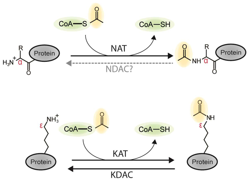
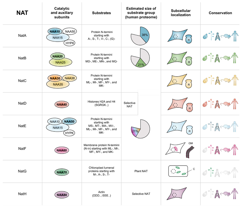
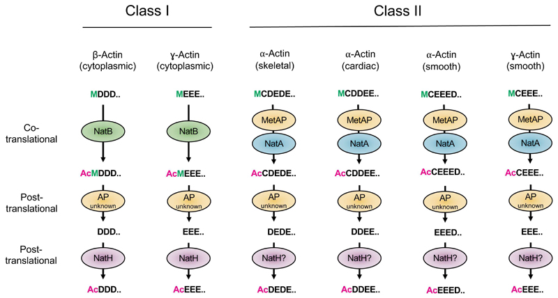
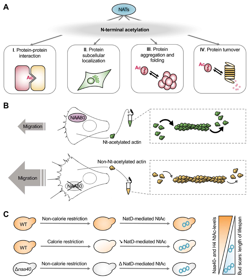
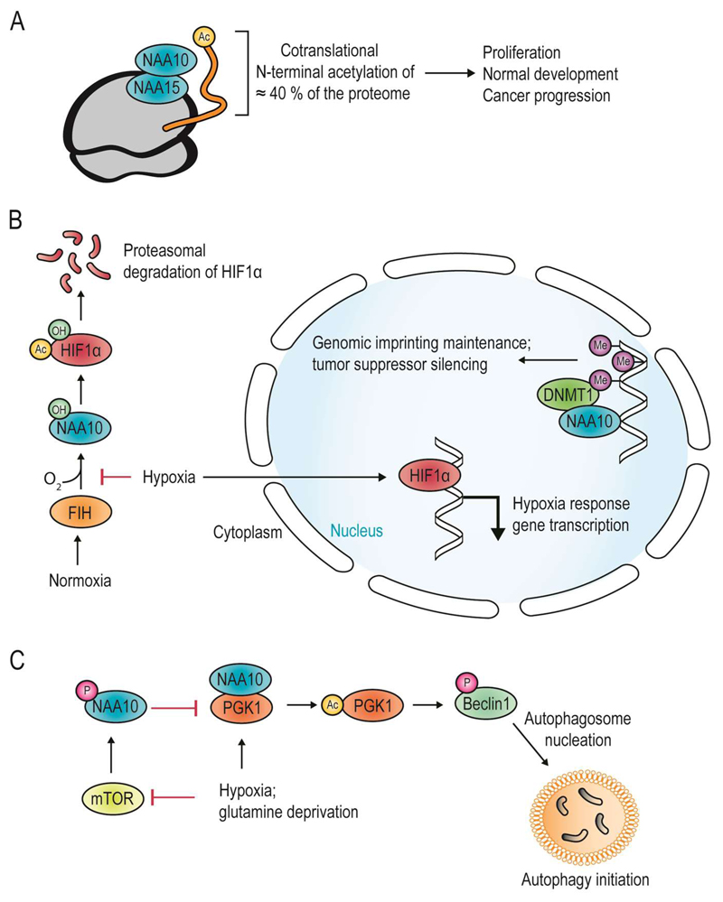
Recent studies of N-terminal acetylation have identified new N-terminal acetyltransferases (NATs) and expanded the known functions of these enzymes beyond their roles as ribosome-associated co-translational modifiers. For instance, the identification of Golgi- and chloroplast-associated NATs shows that acetylation of N termini also happens post-translationally. In addition, we now appreciate that some NATs are highly specific; for example, a dedicated NAT responsible for post-translational N-terminal acetylation of actin was recently revealed. Other studies have extended NAT function beyond Nt acetylation, including functions as lysine acetyltransferases (KATs) and non-catalytic roles. Finally, emerging studies emphasize the physiological relevance of N-terminal acetylation, including roles in calorie-restriction-induced longevity and pathological α-synuclein aggregation in Parkinson's disease. Combined, the NATs rise as multifunctional proteins, and N-terminal acetylation is gaining recognition as a major cellular regulator.

Keywords: KAT; N-terminal acetylation; N-terminal acetyltransferase; NAA10; NAA80; NAT; acetylation; actin; lysine acetyltransferase; protein modifications.


Figures

Figure 1
Figure 1. Protein Nε- and Nα-acetylation.
Protein N-acetyltransferases catalyze the transfer of an acetyl group from acetyl-CoA (green, acetyl group in yellow) onto a substrate protein. An N-terminal acetyltransferase (NAT, top panel) targets the α-amino group (red) of the N-terminal residue, whereas a lysine acetyltransferase (KAT, bottom panel) targets the ε-amine group (red) of an internal lysine. targets. Lysines can be deacetylated by a lysine deacetylase (KDAC), while currently, no N-terminal deacetylases (NDACs) are known, and N-terminal acetylation is thus considered irreversible.
Figure 2
Figure 2. The eukaryotic N-terminal acetyltransferases.
Overview of the composition and characteristics of the eight currently known eukaryotic NATs, NatA to NatH (column 1). The catalytic subunits (NAA10 to NAA80) typically associate with up to two auxiliary subunits (NAA15, NAA25, NAA35, and NAA38) that contribute to activity of the NAT complex through ribosome anchoring and/or substrate specificity-modulation (column 2). NATs have specificity towards different proteins based on the identity of mainly the first two amino acids (column 3) (Aksnes et al., 2016). Taking into account the abundance of each N-termini subtype in the proteome, each NAT is estimated to acetylate a certain percentage of the proteome (column 4) (Aksnes et al., 2016; Aksnes et al., 2015c). NATs vary in their intracellular distribution from cytosolic and ribosome associated (NatA-NatE) to Golgi membrane (NatF), organelle lumen (NatG) and cytosolic but non-ribosomal (NatH) (column 5). In addition, some of the NAT catalytic subunits appear to additionally localize to the nucleus (e.g., NAA10, NAA40 and NAA50; not specifically denoted). The traditional ribosome-associated NATs are well conserved, whereas the more recently discovered NATs performing post-translational Nt-acetylation, are less conserved (column 6). R, Ribosome; GM, Golgi membrane; C, Chloroplast.
Figure 3
Figure 3. N-terminal processing of actin.
In eukaryotes, the Class I actins, β- and γ-actin, match the substrate specificity of NatB and are co-translationally Nt-acetylated by NatB. This acetylated methionine is next cleaved off probably by the enzymatic action of an unidentified aminopeptidase. The newly exposed N-terminus is post-translationally Nt-acetylated by NatH/NAA80. These post-translational steps are specific for animals. The Class II actins, muscle α- and γ-actin, likely undergo co-translational NatA-mediated Nt-acetylation after iMet removal and then follow the same post-translational processing as the Class I actins.
Figure 4
Figure 4. Implications of the N-terminal acetyl group.
(A) A wide spectrum of protein functioning can be affected by Nt-acetylation. Nt-acetylation may be involved in protein-protein interactions. The interaction can directly involve the Nt-acetyl group’s insertion in a hydrophobic binding groove like depicted here or the modification can stabilize a binding region. Correct subcellular localization may be obscured in the absence of Nt-acetylation and in some cases the Nt-acetyl group may stabilize and take part in a membrane binding region. Absence of Nt-acetylation is also associated with aggregation, suggesting a role in global protein folding. In some cases the Nt-acetyl group may act as a proteasomal degradation signal or blocking such a signal, regulating protein lifetime or turnover rate. (B) N-terminal acetylation of actin by NAA80/NatH affects cell morphology and migration rates as well as actin polymerization dynamics in vitro. (C) NatD/Naa40 is downregulated during calorie restriction-induced longevity in yeast and the following reduced Nt-acetylation of histone H4 is suggested to mediate the increased replicative age seen in starved yeast. Deletion of yeast naa40 mimic the effect of calorie restriction.
Figure 5
Figure 5. The diverse catalytic and non-catalytic roles of NAA10.
(A) The classical role of NAA10 as the catalytic subunit of the NatA complex. NatA is found on the ribosome and acetylates approximately 40 % of all nascent N-termini cotranslationally, contributing to diverse cellular functions, such as proliferation, normal organismal development, and as an oncoprotein and potentially a tumor suppressor, depending on the context and the function of the N-terminally acetylated protein. (B) Examples of non-classical roles of NAA10 as a KAT (left) and a non-catalytic regulator (right). Left: NAA10 is hydroxylated during normoxia by factor inhibiting HIF-1α (FIH), which also hydroxylates HIF-1α. Upon hydroxylation, a loop in NAA10 shifts, enabling NAA10 to accommodate lysine substrates and Nε-acetylate HIF-1α. This contributes to the instability and subsequent proteasomal degradation of HIF1α. During hypoxia, neither NAA10 nor HIF-1α are hydroxylated, and HIF-1α is free to translocate to the nucleus and promote transcription of hypoxia response genes. Right: an acetyltransferase-independent function of NAA10. DNA methyltransferase 1 (DNMT1) is recruited to CpG islands by NAA10, which binds to nonmethylated DNA regions. DNMT1 binding to CpG islands furthers genomic imprinting and tumor suppressor silencing. Imprinting is required for normal murine development, but may also contribute to cancer progression. (C) Example of non-classical role of NAA10 as a KAT. NAA10 phosphorylation by mammalian target of rapamycin (mTOR) is inhibited in states of hypoxia or glutamine deprivation. Loss of phosphorylation leads to NAA10 interaction with PGK1 and Nε-acetylation of a PGK1 lysine. Acetylated PGK1 is able to phosphorylate Beclin1 and Beclin1-mediated autophagosome nucleation and autophagy may occur.
Figure 6
Figure 6. Structure of NAA10 and location of pathological mutation sites.
A structural model of NAA10 with SASE peptide substrate and CoA was made in PyMol by aligning the structure of human NAA10 in complex with NAA15 (not shown) (PDB ID: 6C9M; (Gottlieb and Marmorstein, 2018)) to the structure of Schizosaccharomyces pombe NAA10 solved in complex with NAA15 (not shown), CoA and substrate peptide (4KVM; (Liszczak et al., 2013)). CoA is shown as a licorice model colored according to element, SASE substrate peptide is represented in green, CoA-binding residues in yellow and NAA15-binding residues in blue. Mutation sites are indicated with arrows.

References

    1. Akimov V, Barrio-Hernandez I, Hansen SVF, Hallenborg P, Pedersen AK, Bekker-Jensen DB, Puglia M, Christensen SDK, Vanselow JT, Nielsen MM, et al. UbiSite approach for comprehensive mapping of lysine and N-terminal ubiquitination sites. Nature structural & molecular biology. 2018;25:631–640. - PubMed
    1. Aksnes H, Drazic A, Arnesen T. (Hyper)tension release by N-terminal acetylation. Trends in biochemical sciences. 2015a;40:422–424. - PubMed
    1. Aksnes H, Drazic A, Marie M, Arnesen T. First Things First: Vital Protein Marks by N-Terminal Acetyltransferases. Trends in biochemical sciences. 2016;41:746–760. - PubMed
    1. Aksnes H, Goris M, Stromland O, Drazic A, Waheed Q, Reuter N, Arnesen T. Molecular determinants of the N-terminal acetyltransferase Naa60 anchoring to the Golgi membrane. The Journal of biological chemistry. 2017;292:6821–6837. - PMC - PubMed
    1. Aksnes H, Hole K, Arnesen T. Molecular, cellular, and physiological significance of N-terminal acetylation. International review of cell and molecular biology. 2015b;316:267–305. - PubMed

Publication types

Substances