. 2019 Aug:46:29-40.
doi: 10.1016/j.margen.2019.03.003. Epub 2019 Mar 14.

Genomic discovery of ion channel genes in the central nervous system of the lamprey Petromyzon marinus

Affiliations

Genomic discovery of ion channel genes in the central nervous system of the lamprey Petromyzon marinus

Adam J Northcutt et al. Mar Genomics. 2019 Aug.

Abstract

The lamprey is a popular animal model for a number of types of neurobiology studies, including organization and operation of locomotor and respiratory systems, behavioral recovery following spinal cord injury (SCI), cellular and synaptic neurophysiology, comparative neuroanatomy, neuropharmacology, and neurodevelopment. Yet relatively little work has been done on the molecular underpinnings of nervous system function in lamprey. This is due in part to a paucity of gene information for some of the most fundamental proteins involved in neural activity: ion channels. We report here 47 putative ion channel sequences in the central nervous system (CNS) of larval lampreys from the predicted coding sequences (CDS) discovered in the P. marinus genome. These include 32 potassium (K+) channels, six sodium (Na+) channels, and nine calcium (Ca2+) channels. Through RT-PCR, we examined the distribution of these ion channels in the anterior (ARRN), middle (MRRN), and posterior (PRRN) rhombencephalic reticular nuclei, as well as the spinal cord (SC). This study lays the foundation for incorporating more advanced molecular techniques to investigate the role of ion channels in the neural networks of the lamprey.

Keywords: Genome; Ion channel; Reticulospinal.


Conflict of interest statement

Conflict of Interest Statement

The authors declare that they have no competing interests.

Figures
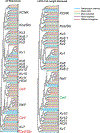
Figure 1.
Figure 1.. COBALT cluster dendrogram analysis of identified P. marinus ion channel sequences.
P. marinus (Pm-) ion channel amino acid sequences were used in an analysis with orthologous Danio rerio, Mus musculus, Homo sapiens, and Rattus norvegicus ion channel amino acid sequences to create a cluster dendrogram using NCBI’s COBALT (left). Ion channel families are labeled, and species identification is colored with P. marinus = cyan, D. rerio = orange, M. musculus = green, H. sapiens = purple, R. norvegicus = red. The red lettering indicates group identifications that were split across multiple nodes, and suggested discrepancies in the ability of COBALT to appropriately resolve relationships. As a result, we systematically removed sequences from the analysis based on their length, and reiterated the clustering until more coherence was achieved in the grouping of channel families (right dendrogram). Green lettering indicates group identification that was improved following removal of sequences with short predicted length (<25% full length). Red arrows indicate the following four P. marinus sequences that were removed between the analyses on the left and right dendrograms (Pm- Kv7c, Pm-Cav1b, Pm-Cav2a, and Pm-Cav2c).
Figure 2.
Figure 2.. Two-pore-domain Potassium (KCNK) and Calcium/Sodium-Activated Potassium Channels (Slo and Kca) identified in P. marinus.
We used the clustering from the combined COBALT analysis in Figure 1 to subset channel families for more detailed phylogenetic analysis. Because the KCNK and Slo/Kca channels formed a coherent node in the combined analysis, we performed subsequent (A) COBALT as well as (B) Minimum Evolution (ME) and (C) Neighbor Joining (NJ) phylogenetic analyses on these predicted amino acid sequences from putative lamprey ortholog coding sequences as well as protein sequences from Mus musculus (Mm) and Danio rerio (Dr). Lamprey (Petromyzon marinus, Pm) sequence is noted in red. This analysis included a previously identified SKKCa sequence, labeled as Pm-SKKCa** (McClellan et al. 2008). All putative ortholog sequences from lamprey are included in the COBALT analysis, while ME and NJ results in some sequences not being retained due to exceeding the maximum allowed fraction of mismatched bases in the aligned sequences.
Figure 3.
Figure 3.. Voltage-gated (Kv) potassium channel families and subtypes identified in P. marinus.
Voltage-gated K+ channels of the (A-C) Kv1, Kv2, Kv3, Kv5, Kv6, Kv8 and Kv9 families are represented based on their initial clustering patterns in the combined COBALT analysis in Figure 1. No Kv4 type channels were identified by BLAST analysis. Plots were generated and are presented in the same manner as described in Figure 2.
Figure 4.
Figure 4.. Eag, HCN, and KCNQ/M-type channel families and subtypes identified in P. marinus.
Channels of the (A-C) Kv10–12 (Eag-like), HCN, and (D-F) Kv7 (KCNQ/M-type) families are represented based on their initial clustering patterns in the combined COBALT analysis in Figure 1. Plots were generated and are presented in the same manner as described in Figure 2.
Figure 5.
Figure 5.. Voltage-gated sodium and calcium channel families and subtypes identified in P. marinus.
Voltage-gated Na+ and Ca2+ channels of the (A-C) Nav and Cav1, Cav2, and Cav3 families are represented based on their initial clustering patterns in the combined COBALT analysis in Figure 1. Trees were generated in the same manner as described in Figure 2. This analysis included a previously identified high-voltage activated (HVA) calcium channel sequence, labeled as Pm-HVA-Calcium** (McClellan et al., 2008). Sodium channels are named based on work that initially described these proteins in lamprey (Zakon et al., 2017).
Figure 6.
Figure 6.. P. marinus central nervous system ion channel distribution.
(A) Diagram of lamprey brain (left) and upper spinal cord (SC). Contours showing the four reticular nuclei (Shaw et al., 2010): mesencephalic reticular nucleus (MRN) and anterior (ARRN), middle (MRRN), and posterior (PRRN) rhombencephalic reticular nuclei. Dotted lines indicate tissues collected for RNA extraction and subsequent PCR reactions (see Methods). (B) Images of 2% agarose gels stained with ethidium bromide that were used to resolve gene-specific amplicons generated from PCR reactions on mixed brain and spinal cord tissues. The first lane of each row contained GeneRuler 100 bp ladder with the 500 bp sequence indicated with an arrow. Assay 1 was designed with amplicons around 500 bp (see Table 2), while Assay2 was designed with amplicons around 300 bp (see Table 3).

Similar articles

Cited by

References

    1. Alford ST, Alpert MH, 2014. A synaptic mechanism for network synchrony. Front. Cell. Neurosci 8, 290. doi:10.3389/fncel.2014.00290 - DOI - PMC - PubMed
    1. Batueva IV, Tsvetkov EA, Buchanan JT, Veselkin NP, 1996. Investigation of voltage- gated currents in the isolated neurons of river lamprey Lampetra fluviatilis spinal cord. J Evol 217–231. - PubMed
    1. Benarroch EE, 2013. HCN channels: Function and clinical implications. Neurology 80, 304–310. doi:10.1212/WNL.0b013e31827dec42 - DOI - PubMed
    1. Cantarel BL, Korf I, Robb SMC, Parra G, Ross E, Moore B, Holt C, Alvarado AS, Yandell M, 2008. MAKER: An easy-to-use annotation pipeline designed for emerging model organism genomes. Genome Res 18, 188–196. doi:10.1101/gr.6743907 - DOI - PMC - PubMed
    1. Catterall WA, 2005. International Union of Pharmacology. XLVII. Nomenclature and Structure- Function Relationships of Voltage-Gated Sodium Channels. Pharmacol. Rev 57, 397–409. doi:10.1124/pr.57.4.4 - DOI - PubMed

LinkOut - more resources