Review
. 2019 Aug:57:93-102.
doi: 10.1016/j.sbi.2019.02.008. Epub 2019 Mar 14.

Advances in high-speed atomic force microscopy (HS-AFM) reveal dynamics of transmembrane channels and transporters

Affiliations
Review

Advances in high-speed atomic force microscopy (HS-AFM) reveal dynamics of transmembrane channels and transporters

George R Heath et al. Curr Opin Struct Biol. 2019 Aug.

Abstract

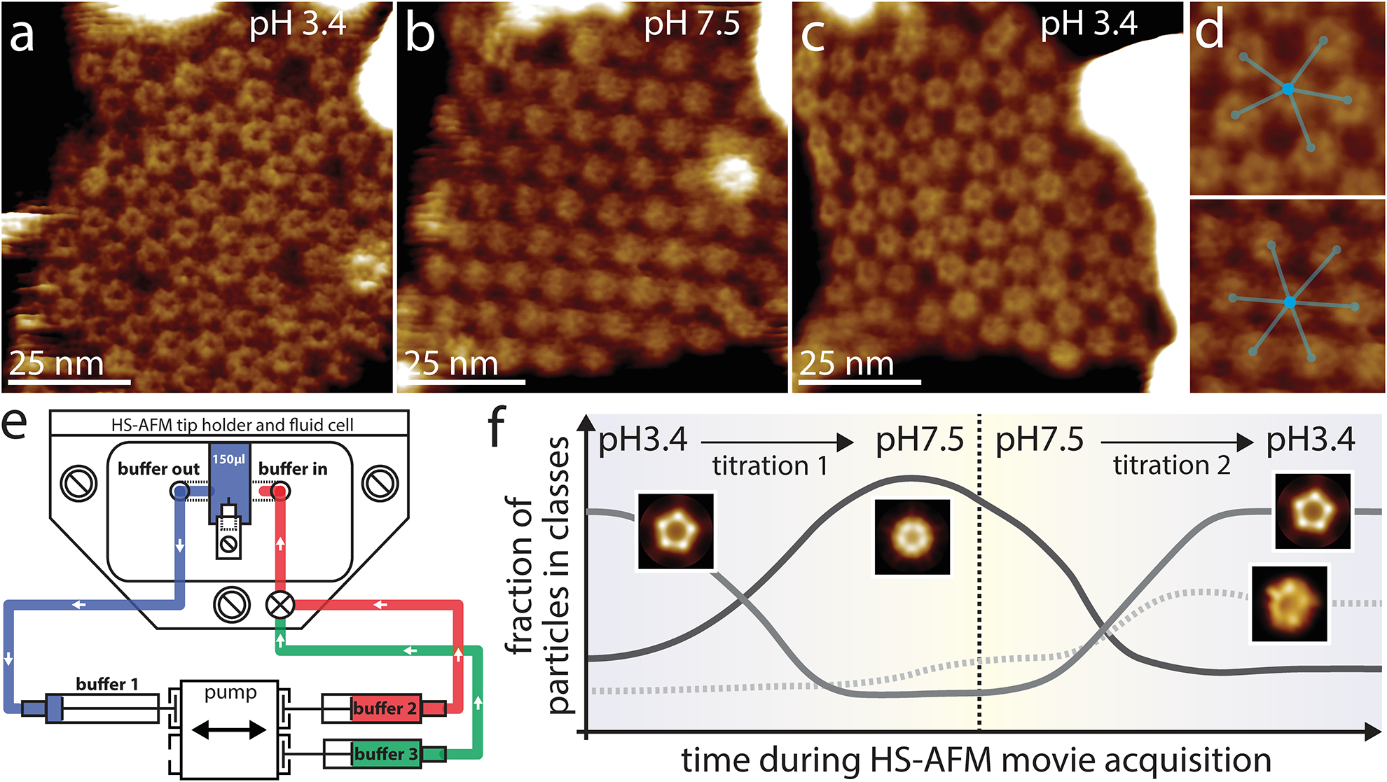
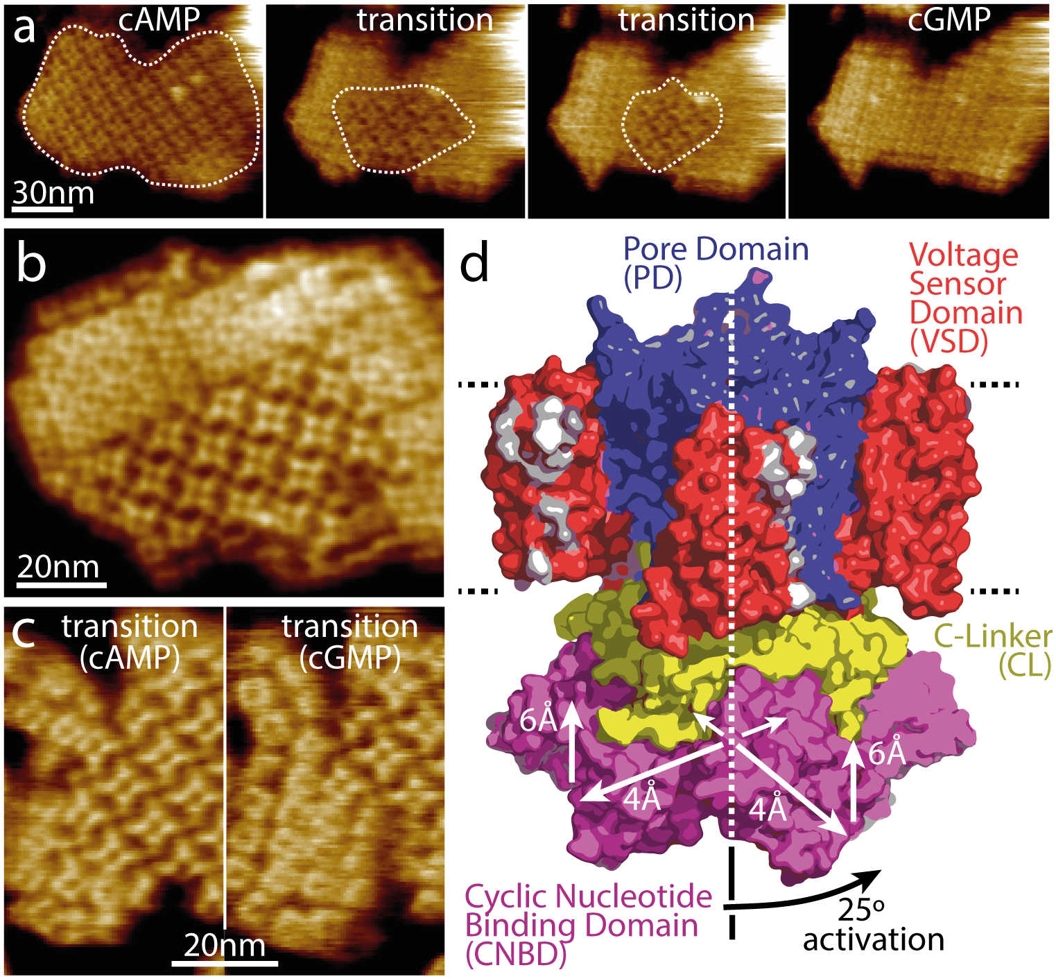
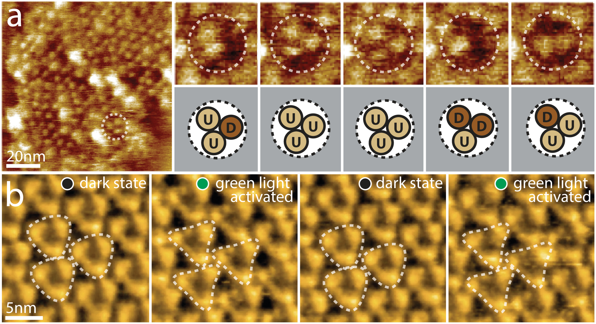
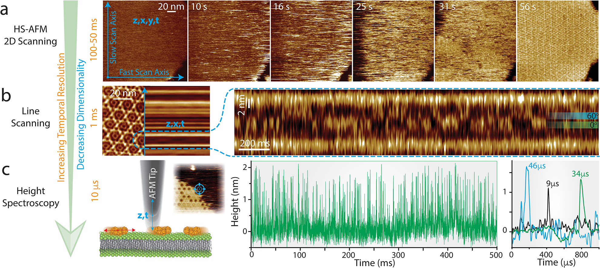
Recent advances in high-speed atomic force microscopy (HS-AFM) have made it possible to study the conformational dynamics of single unlabeled transmembrane channels and transporters. Improving environmental control with the integration of a non-disturbing buffer exchange system, which in turn allows the gradual change of conditions during HS-AFM operation, has provided a breakthrough toward the performance of structural titration experiments. Further advancements in temporal resolution with the use of line scanning and height spectroscopy techniques show how high-speed atomic force microscopy can measure millisecond to microsecond dynamics, pushing this method beyond current spatial and temporal limits offered by less direct techniques.


Conflict of interest statement

Conflict of interest statement

Nothing declared.

Figures

Figure 1.
Figure 1.. HS-AFM structural titration experiments of GLIC channels.
During HS-AFM operation, the pH in the fluid chamber was gradually exchanged through a buffer exchange microfluidic device from pH 3.4 (a) to pH 7.5 (b) and back to pH 3.4 (c). Analysis of the supramolecular assemblies show reversible rearrangements from seemingly disordered arrangements, where channels assemble in trimers of pentamers and most channels have 5 neighbors (yellow outlines), to hexagonal packing and back again (the white dashed outlines and the inset in (c) highlight higher-order structures in the different conditions). (d) Zoom-in of the molecular arrangements at pH3.4 (top) and pH7.5 (bottom) with ball/stick overlays to highlight how 5 neighbors are preferred at low pH over 6 at higher pH. (e) Schematic representation of the coupling of the buffer exchange system to the HS-AFM fluid cell. The ‘buffer in’ and ‘buffer out’ channels are placed in proximity to the HS-AFM cantilever (central element inside the fluid cell). The fluid cell contains initially 150ul of buffer 1 (blue) that is connected to the receiving syringe. Buffer 2 (red) is the first buffer to be injected into the fluid cell. Note the slight gap between buffer 2 and the fluid cell, representing a tiny bubble in the tubing to avoid involuntary mixing of buffer 2 before activation of the pumping system. By the use of a switch in the tubing system, buffer 3 (green) can later be injected into the fluid cell. Buffer 3 can be identical to buffer 1, allowing for a reversibility experiment. (f) Abundance of GLIC channel conformations as a function of time as determined by reference-free 2D classification. At the beginning and end of the movie, at pH 3.4, the active/desensitized-state, a class where the ECDs form a flower-shaped pentamer surrounding a central cavity are most abundant. In the middle of the movie, at pH 7.5, the closed-state conditions, a class with narrowed ECDs dominates. An asymmetric class exists throughout the experiment, but peaks upon re-exposure to low activating pH.
Figure 2.
Figure 2.. Dynamics of ligand-induced conformational changes in SthK by real-time HS-AFM imaging.
(a) HS-AFM time-lapse high-resolution image sequence of a SthK 2D-crystal initially in 0.1mM cAMP and exposing CNBDs. Upon addition of 7mM cGMP, SthK channels undergo a conformational change progressively from the borders to the center of the membrane patch (dotted outline). (b) High-resolution topography of a membrane containing well-ordered channels in both conformations. (c) High-resolution topographs during a cAMP to cGMP transition where the majority of the molecules are in the cAMP (left) and in the cGMP (right) conformation, respectively. (d) Model of SthK in the activated state: Upon activation, the CNBDs rotate by ~25° clockwise (when viewed from the intracellular side) and move by ~6 Å towards the membrane and by ~4 Å outwards from the four-fold axis (note, this activated state illustration is a cartoon using domains of the SthK structure repositioned according to the displacements found by HS-AFM).
Figure 3.
Figure 3.. Active domain motions in the glutamate transporter GltPh and the light-driven proton pump bR
(a) Direct visualization of GltPh transport domain elevator movements by HS-AFM. Left: A typical HS-AFM image of a GltPh reconstituted membrane displays densely packed GltPh trimers (dashed outline). Right: Conformational dynamics of a representative GltPh trimer under substrate-free conditions (imaging rate: 1 s−1, frame size: 20nm). Each GltPh protomer in the trimer (top) shows reversible conformational alternation between outward facing (up, U) and inward facing (down, D) states (bottom). (b) HS-AFM movie frames of D96N bacteriorhodopsin (bR) exposed to repeated dark and green light illumination cycles (imaging rate: 1 s−1). bR trimers are highlighted by the white dashed triangles. Under illumination, conformational changes result in significant changes in the topography, notably a movement of the E-F loop outwards from the 3-fold axis (E-F loops of neighboring activated trimers interact closely in the activated state).
Figure 4.
Figure 4.. HS-AFM line scanning (HS-AFM-LS) and HS-AFM height spectroscopy (HS-AFM-HS): Increasing the temporal resolution by reducing the dimensionality of data acquisition.
(a) HS-AFM 2D-scanning movie of A5 membrane-binding, self-assembly and formation of p6 2D-crystals upon UV-illumination-induced Ca2+-release. Blue arrows illustrate the slow (vertical) and the fast (horizontal) scan axes. Images can be captured at up to 10–20 images per second. (b) Left: averaged HS-AFM image of an A5 p6-lattice overlaid with the subsequent line scanning kymograph, obtained by scanning repeatedly the central x-direction line as illustrated by the blue arrow with a maximum rate of 1000–2000 lines per second. Right: line scanning kymograph across one protomer of the non-p6 trimer, outlined by the semicircular dashed line in the 2D image (left), at a rate of 417 lines per second (2.4 ms/line). Overlaid on the kymography are the positions of the 0° and 60° states that the trimer alternately adopts. (c) Left: schematic showing the principle of HS-AFM height spectroscopy (HS-AFM-HS) allowing 10 μs temporal resolution. The AFM tip is oscillated in z at a fixed x,y-position, detecting single molecule dynamics such as diffusion under the tip (inset: HS-AFM image of an A5 p6-lattice partially covering the membrane surface during self-assembly, HS-AFM-HS is performed at a fixed position at the center of the image as illustrated by the target). Right: Height/time traces obtained by HS-AFM-HS allowing determination of the local A5 concentration and diffusion rates (the colored traces on the very right are a zoomed overlay of three traces displaying diffusion events under the tip in the low microsecond time range).

References

    1. Scheres SHW: RELION: Implementation of a Bayesian approach to cryo-EM structure determination. J Struct Biol 2012, 180:519530. - PMC - PubMed
    1. Liao M, Cao E, Julius D, Cheng Y: Structure of the TRPV1 ion channel determined by electron cryo-microscopy. Nature 2013, 504:107–112. - PMC - PubMed
    1. Cheng Y: Membrane protein structural biology in the era of single particle cryo-EM. Curr Opin Struct Biol 2018, 52:58–63. - PMC - PubMed
    1. Drew D, Boudker O: Shared Molecular Mechanisms of Membrane Transporters. Annu Rev Biochem 2016, 85:543–572. - PubMed
    1. Bavi N, Martinac AD, Cortes DM, Bavi O, Ridone P, Nomura T, Hill AP, Martinac B, Perozo E: Structural Dynamics of the MscL C-terminal Domain. Sci Rep 2017, 7:17229. - PMC - PubMed

MeSH terms

Substances