

Localized multiple malignant epithelioid peritoneal mesotheliomas arising from the hepatoduodenal ligament and diaphragm: a case report
- PMID: 30879467
- PMCID: PMC6421649
- DOI: 10.1186/s13256-019-2008-9
Localized multiple malignant epithelioid peritoneal mesotheliomas arising from the hepatoduodenal ligament and diaphragm: a case report
Abstract
Background: Malignant peritoneal mesothelioma is a rare aggressive tumor of the peritoneum. We report a rare case of resection of multiple localized malignant peritoneal mesotheliomas.
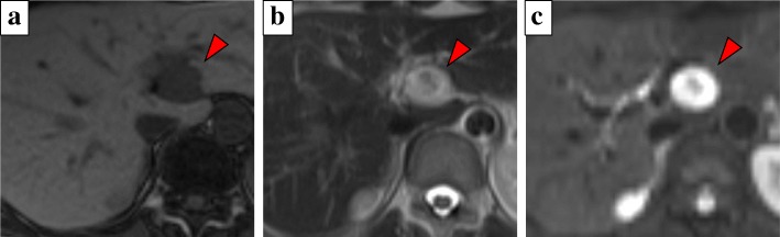
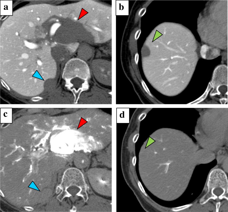
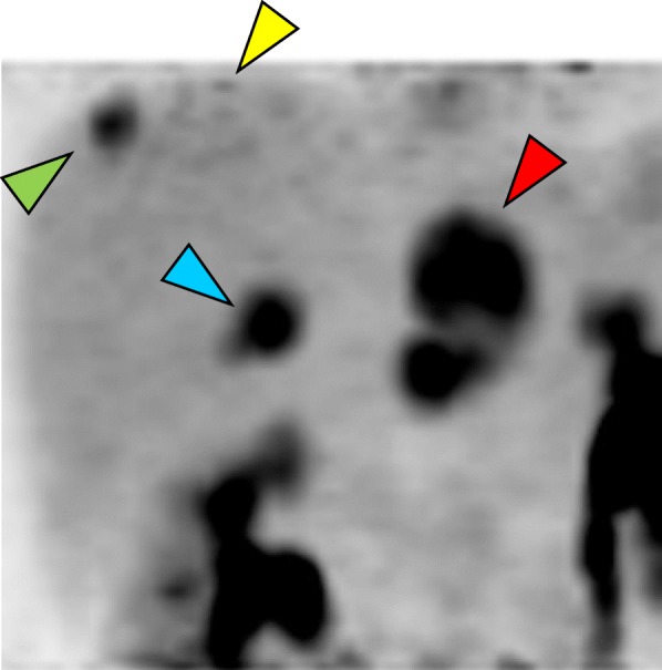
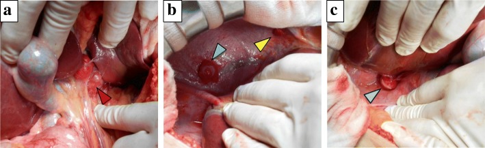
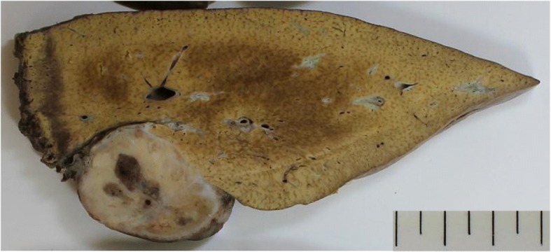
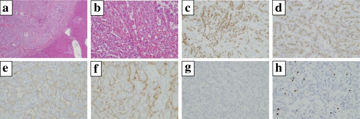
Case presentation: A 55-year-old Japanese woman was admitted to our hospital because liver tumors were detected by abdominal ultrasonography during a screening examination. Blood examination findings, including tumor makers, were within normal ranges. She had no evidence of exposure to asbestos. Computed tomography showed four hypervascular, round liver tumors, one in the lateral liver segment adjacent to the hepatic hilus, and the other three on the liver surface. Computed tomography angiography revealed that the tumor in the lateral segment had strong enhancement and was fed from the left gastric artery. In contrast, the other tumors showed no enhancement, and were fed from the right inferior phrenic artery. Abnormal accumulation was identified in the four tumors only with 18F-fluorodeoxyglucose positron emission tomography. It was very difficult to obtain a definitive preoperative diagnosis, but surgical resection was performed because we considered potential malignancy. Laparotomy revealed the principal site of the tumor in the lateral segment was on the hepatoduodenal ligament, and all other tumors were on the diaphragm. A left lobectomy and partial diaphragmatic resection were performed. The final pathological diagnosis was multiple malignant epithelioid mesotheliomas. Our patient has had no recurrence for 20 months postoperatively.
Conclusions: In general, malignant peritoneal mesotheliomas are classified as diffuse tumors, which are often unresectable and have a poor prognosis. However, early diagnosis and treatment, particularly with the localized type, as in our patient, could lead to long-term survival of the patient. We recommend that multiple malignant epithelioid mesotheliomas be included in the differential diagnosis for patients with subcapsular hepatic tumors.
Keywords: Angiography; Case report; Localized mesothelioma; Malignant peritoneal mesothelioma.
Conflict of interest statement
Ethics approval and consent to participate
Not applicable.
Consent for publication
Written informed consent was obtained from the patient for publication of this case report and any accompanying images. A copy of the written consent is available for review by the Editor-in-Chief of this journal.
Competing interests
The authors declare that they have no competing interests.
Publisher’s Note
Springer Nature remains neutral with regard to jurisdictional claims in published maps and institutional affiliations.
Figures

Similar articles
-
Primary malignant mesothelioma of the diaphragm with liver invasion: A case report and review of literature.Medicine (Baltimore). 2019 Apr;98(15):e15147. doi: 10.1097/MD.0000000000015147. Medicine (Baltimore). 2019. PMID: 30985689 Free PMC article. Review.
-
Multiple primary hepatic malignant mesotheliomas mimicking cystadenocarcinomas on enhanced CT and FDG PET/CT.Clin Nucl Med. 2014 Jul;39(7):619-22. doi: 10.1097/RLU.0b013e31828da61d. Clin Nucl Med. 2014. PMID: 23510896
-
Peritoneal Deciduoid Malignant Mesothelioma on 67Ga SPECT/CT.Clin Nucl Med. 2019 Feb;44(2):161-163. doi: 10.1097/RLU.0000000000002363. Clin Nucl Med. 2019. PMID: 30394926
-
A Case of Localized Malignant Peritoneal Mesothelioma Evaluated by 18F-FDG PET/CT.Clin Nucl Med. 2020 Nov;45(11):890-891. doi: 10.1097/RLU.0000000000003158. Clin Nucl Med. 2020. PMID: 32604114
-
Diffuse malignant epithelioid mesothelioma in a background of benign multicystic peritoneal mesothelioma: a case report and review of the literature.BMJ Case Rep. 2014 Feb 5;2014:bcr2013200212. doi: 10.1136/bcr-2013-200212. BMJ Case Rep. 2014. PMID: 24501333 Free PMC article. Review.
Cited by
-
Recurrent malignant peritoneal mesothelioma treated by a second resection: A case report.Clin Case Rep. 2023 May 18;11(5):e7383. doi: 10.1002/ccr3.7383. eCollection 2023 May. Clin Case Rep. 2023. PMID: 37215970 Free PMC article.
-
Primary Intrahepatic Mesothelioma: Case Series and Systematic Review of Literature.J Gastrointest Cancer. 2024 Dec;55(4):1520-1529. doi: 10.1007/s12029-024-01075-x. Epub 2024 Aug 14. J Gastrointest Cancer. 2024. PMID: 39141212
References
Publication types
MeSH terms
LinkOut - more resources
Full Text Sources
Medical
