. 2019 May 16;26(5):699-710.e6.
doi: 10.1016/j.chembiol.2019.02.007. Epub 2019 Mar 14.

Tracing MYC Expression for Small Molecule Discovery

Affiliations

Tracing MYC Expression for Small Molecule Discovery

Jutta Steinberger et al. Cell Chem Biol. .

Abstract

Our inability to effectively "drug" targets such as MYC for therapeutic purposes requires the development of new approaches. We report on the implementation of a phenotype-based assay for monitoring MYC expression in multiple myeloma cells. The open reading frame (ORF) encoding an unstable variant of GFP was engineered immediately downstream of the MYC ORF using CRISPR/Cas9, resulting in co-expression of both proteins from the endogenous MYC locus. Using fluorescence readout as a surrogate for MYC expression, we implemented a pilot screen in which ∼10,000 compounds were prosecuted. Among known MYC expression inhibitors, we identified cardiac glycosides and cytoskeletal disruptors to be quite potent. We demonstrate the power of CRISPR/Cas9 engineering in establishing phenotype-based assays to identify gene expression modulators.

Keywords: CRISPR/Cas9; MYC expression; chemical biology; drug discovery; drug repurposing; genome engineering; multiple myelomas; phenotype-based assay; target identification.


Conflict of interest statement

DECLARATION OF INTERESTS

The authors declare no competing financial interests.

Figures

Figure 1.
Figure 1.
Tracing MYC expression with CRISPR/Cas9 engineered JJN-3 cells. See also Figures S1 and S2. A. Schematic diagram of the MYC locus. Two promoters (P1, P2) and three translation initiation sites are denoted. The locations of the PAM, sgRNA and MYC termination codon (TAA: asterisk) are shown. The black triangle denotes the predicted Cas9 cleavage site. Recombination of the MYCP2A/d2GFP template by HDR will lead to an in-frame insertion of the P2A/d2GFP fusion ORF immediately downstream of the MYC ORF. The red triangle indicates the location of P2A cleavage site. B. Northern blot analysis of RNA from JJN-3 and D11 cells probed with a DNA fragment spanning the MYC 3’ UTR or the GFP ORF. C. Western blot analysis of protein extracts prepared from JJN-3 and D11 cells. Antibodies used for probings are indicated to the left of the panels.
Figure 2.
Figure 2.
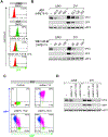
Phenotypic response of D11 cells to MYC expression inhibitors. A. Flow cytometry analysis of JJN-3 and D11 cells exposed to the BET bromodomain inhibitor JQ1 or the translation inhibitor CR-1–31-B. The % GFP+ gated cells is indicated in the top right quadrant. B. Western blot analysis for MYC and GFP in JJN-3 and D11 cells subjected to the indicated treatments. C. Flow cytometry analysis of D11 cells infected with pPRIME/BFP expressing a neutral control shRNA to Renilla luciferase (shRen.713) or shRNAs targeting MYC (shMyc.1702 and shMyc.1831). D. Western blot analysis of extracts from JJN-3 and D11 cells infected with pPRIME/BFP expressing the indicated shRNAs. -, no infection.
Figure 3.
Figure 3.
HTS Prosecution. See also Figures S3 and S6 and Tables S1 and S2. A. Results from screening an FDA approved drug collection, an in-house curated NP repository, and a bioactive compound set. The dotted line represents the 3 x SD cut-off. The blue dots represent spiked-in positive controls (50 nM CR-1–31-B) and red dots represent hits. Z factor = 0.78. B. Strategy used to generate JJN-3/d2GFP cells which were used in counter-screens. C. A comparison of the response of D11 and JJN-3/d2GFP to the indicated compounds. The proportion of GFPhigh cells was determined by flow cytometry and is indicated in the top right quadrant. D. Heat map displaying results from the counter-screen assay using JJN-3/d2GFP cells compared to those obtained with the D11 line. Raw values are presented in Table S2. Compounds in red text show no selectivity between D11 and JJN-3/d2GFP, those in blue are known inhibitors of translation, and green text indicates cardiac glycosides.
Figure 4.
Figure 4.
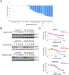
Target engagement of the Na+/K+ ATPase is required for CG activity towards MYC. See also Figure S4 and Table S3. A. Waterfall plot showing percent change from baseline (GFP+ to GFP) in D11 cells following exposure for 5 h to the indicated bufadienolides (see Table S3 for structures). n=2, ±SEM. B. JJN-3 cells expressing FF or mAtp1a1 cDNA were exposed to the indicated concentrations of bufalin, ouabain, or silvestrol for 5 h after which cell extracts were prepared and probed by Western blotting using the indicated antibodies. C. Viability of JJN-3 cells expressing the FF or mAtp1a1 cDNA when exposed to bufalin, silvestrol, or ouabain for 48 h. n=2 ± SD.
Figure 5.
Figure 5.
Effects of CGs on MYC transcription and translation. See also Figures S4 and S5. A. Quantitation of MYC mRNA levels in JJN-3 cells treated with the indicated compounds for 5 h. RNA was isolated from cells and analyzed by RT-qPCR. MYC mRNA levels are normalized to actin mRNA levels. Compounds were used at the following concentrations: ouabain (125 nM), bufalin (50 nM), proscillaridin A (50 nM), digoxin (250 nM), and digitoxin (125 nM). B. Kinetic analysis of MYC expression and induction of eIF2α phosphorylation following bufalin treatment. C. JJN-3 cells were incubated in the presence of the indicated compounds (50 nM) for 1 and 5 h after which time 35S-methionine was added to the media for another 15 mins. Cells were then harvested, lysed, and radioactive protein levels measured by TCA precipitation followed by scintillation counting. n=3, ± SD. D. Cytotoxicity of bufalin towards hematological cancer cells. IC50’s were determined from a 10 point titration. Cells were incubated with compound for 48 h, after which time cell viability was determined by Cell Titer-Glo. n=3, ± SD.
Figure 6.
Figure 6.
Bioassay guided fractionation of natural product extracts uncovers cytoskeleton targeting molecules. See also Figure S6. A. Structure of jasplakinolide. B. Structure of dolastatin-12. C. Quantitation of MYC, eEF1α, eEF2, and GADPH mRNA levels in JJN-3 cells treated with the indicated compounds for 5 h (JQ1 [250 nM], jasplakinolide [250 nM], dolastatin-12 [1 μM]). MYC mRNA levels are relative to levels obtained in the absence of compound. n=3 ± SD. D. JJN-3 cells were incubated 5 h in the presence of the indicated compounds, at which point extracts were prepared, fractionated by SDS-PAGE, transferred to PVDF membrane, and probed with antibodies to the indicated proteins. Concentrations used were: dolastatin-12 (16, 64, 256 nM), jasplakinolide (32, 128, 512 nM), latruncullin (310, 1250, 5000 μM), nocodazole (0.625, 2.5, 10 uμM), swinhollide (40, 156, 625 nM), colcemid (0.625, 2.5, 10 μM), phalloidin (0.625, 2.5, 10 μM), paclitaxel (0.625, 2.5, 10 μM), vinblastine (0.625, 2.5, 10 μM), colchicine (0.625, 2.5, 10 μM), and vincristine (0.625, 2.5, 10 μM).

References

    1. Beroukhim R, Mermel CH, Porter D, Wei G, Raychaudhuri S, Donovan J, Barretina J, Boehm JS, Dobson J, Urashima M, et al. (2010). The landscape of somatic copy-number alteration across human cancers. Nature 463, 899–905. - PMC - PubMed
    1. Bhat M, Robichaud N, Hulea L, Sonenberg N, Pelletier J, and Topisirovic I (2015). Targeting the translation machinery in cancer. Nat Rev Drug Discov 14, 261–278. - PubMed
    1. Bouvard C, Lim SM, Ludka J, Yazdani N, Woods AK, Chatterjee AK, Schultz PG, and Zhu S (2017). Small molecule selectively suppresses MYC transcription in cancer cells. Proc Natl Acad Sci USA 114, 3497–3502. - PMC - PubMed
    1. Braet F, Spector I, De Zanger R, and Wisse E (1998). A novel structure involved in the formation of liver endothelial cell fenestrae revealed by using the actin inhibitor misakinolide. Proc Natl Acad Sci USA 95, 13635–13640. - PMC - PubMed
    1. Brock A, Huang S, and Ingber DE (2003). Identification of a distinct class of cytoskeleton-associated mRNAs using microarray technology. BMC Cell Biol 4, 6. - PMC - PubMed

Publication types

MeSH terms

LinkOut - more resources