

Elastic fibers and biomechanics of the aorta: Insights from mouse studies
- PMID: 30880160
- PMCID: PMC6745291
- DOI: 10.1016/j.matbio.2019.03.001
Elastic fibers and biomechanics of the aorta: Insights from mouse studies
Abstract
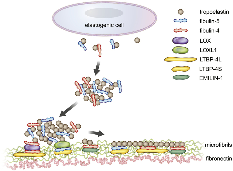
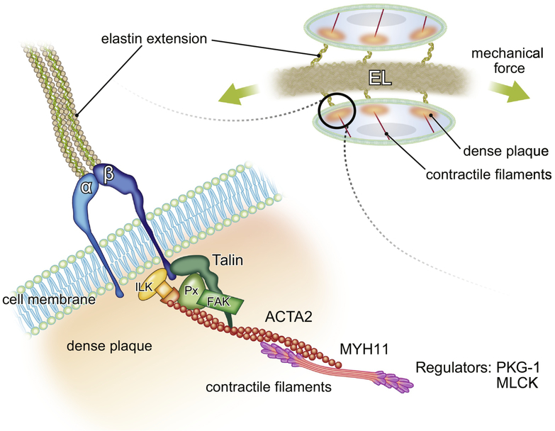
Elastic fibers are major components of the extracellular matrix (ECM) in the aorta and support a life-long cycling of stretch and recoil. Elastic fibers are formed from mid-gestation throughout early postnatal development and the synthesis is regulated at multiple steps, including coacervation, deposition, cross-linking, and assembly of insoluble elastin onto microfibril scaffolds. To date, more than 30 molecules have been shown to associate with elastic fibers and some of them play a critical role in the formation and maintenance of elastic fibers in vivo. Because the aorta is subjected to high pressure from the left ventricle, elasticity of the aorta provides the Windkessel effect and maintains stable blood flow to distal organs throughout the cardiac cycle. Disruption of elastic fibers due to congenital defects, inflammation, or aging dramatically reduces aortic elasticity and affects overall vessel mechanics. Another important component in the aorta is the vascular smooth muscle cells (SMCs). Elastic fibers and SMCs alternate to create a highly organized medial layer within the aortic wall. The physical connections between elastic fibers and SMCs form the elastin-contractile units and maintain cytoskeletal organization and proper responses of SMCs to mechanical strain. In this review, we revisit the components of elastic fibers and their roles in elastogenesis and how a loss of each component affects biomechanics of the aorta. Finally, we discuss the significance of elastin-contractile units in the maintenance of SMC function based on knowledge obtained from mouse models of human disease.
Copyright © 2019 Elsevier B.V. All rights reserved.
Figures
References
-
- Safar ME, Levy BI, Struijker-Boudier H (2003). Current perspectives on arterial stiffness and pulse pressure in hypertension and cardiovascular diseases. Circulation 107, 2864–2869. - PubMed
-
- Davis EC (1993). Smooth muscle cell to elastic lamina connections in developing mouse aorta. Role in aortic medial organization. Lab Invest 68, 89–99. - PubMed
-
- Nakamura T, Lozano PR, Ikeda Y, Iwanaga Y, Hinek A, Minamisawa S, Cheng CF, Kobuke K, Dalton N, Takada Y, Tashiro K, Ross J Jr., Honjo T, Chien KR (2002). Fibulin-5/DANCE is essential for elastogenesis in vivo. Nature 415, 171–175. - PubMed
-
- Yanagisawa H, Davis EC, Starcher BC, Ouchi T, Yanagisawa M, Richardson JA, Olson EN (2002). Fibulin-5 is an elastin-binding protein essential for elastic fibre development in vivo. Nature 415, 168–171. - PubMed
Publication types
MeSH terms
Substances
Grants and funding
LinkOut - more resources
Full Text Sources
