. 2019 Mar 18;11(2):12.
doi: 10.1038/s41368-019-0046-1.

Overexpression of Dlx2 enhances osteogenic differentiation of BMSCs and MC3T3-E1 cells via direct upregulation of Osteocalcin and Alp

Affiliations

Overexpression of Dlx2 enhances osteogenic differentiation of BMSCs and MC3T3-E1 cells via direct upregulation of Osteocalcin and Alp

Jianfei Zhang et al. Int J Oral Sci. .

Abstract

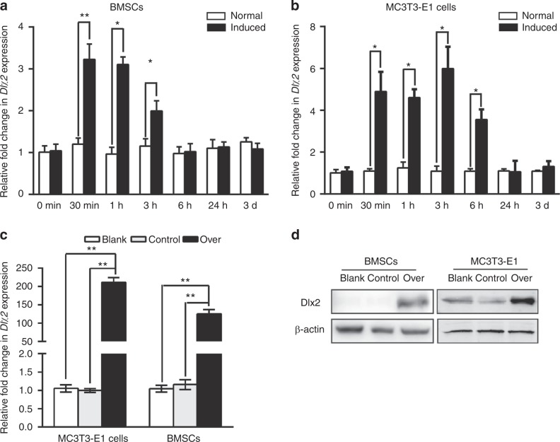
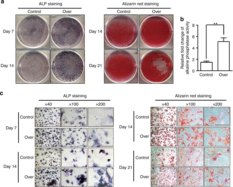
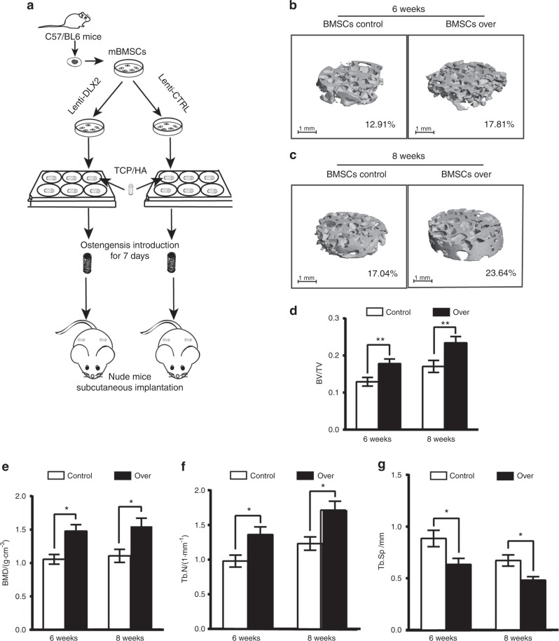
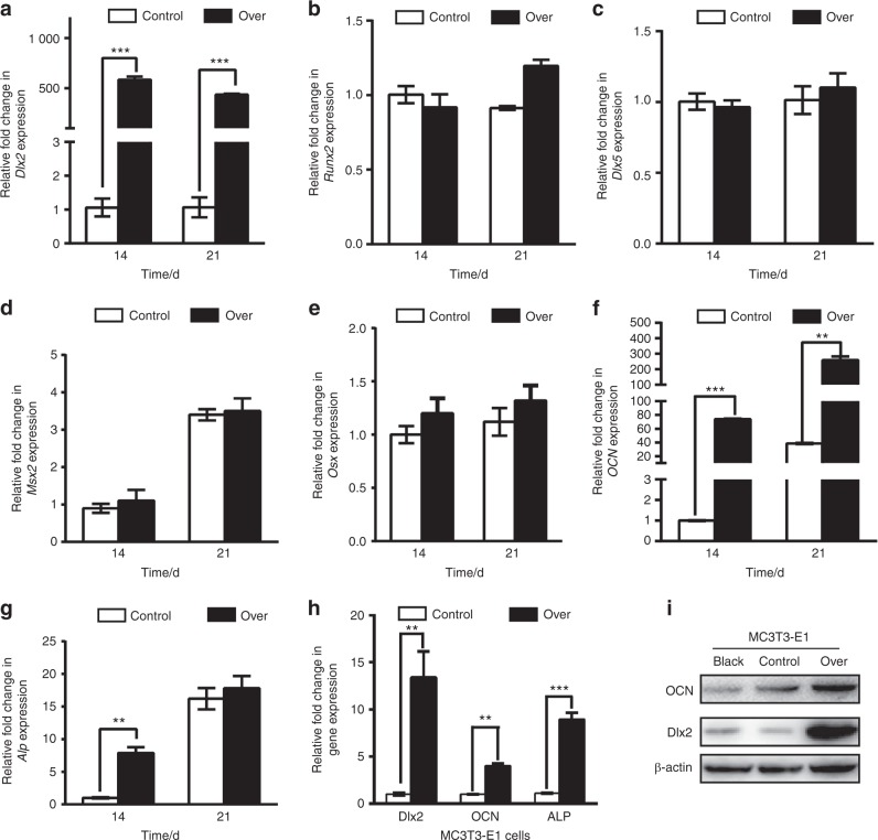
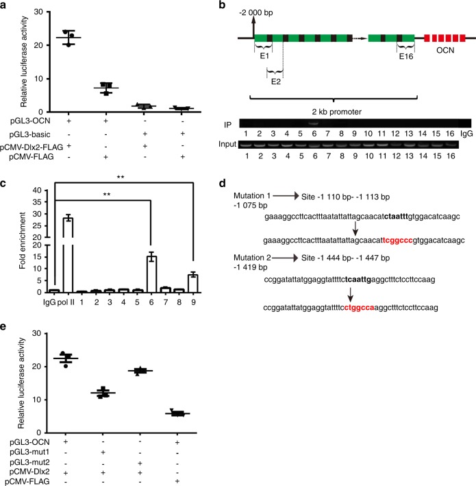
Genetic studies have revealed a critical role of Distal-homeobox (Dlx) genes in bone formation, and our previous study showed that Dlx2 overexpressing in neural crest cells leads to profound abnormalities of the craniofacial tissues. The aim of this study was to investigate the role and the underlying molecular mechanisms of Dlx2 in osteogenic differentiation of mouse bone marrow stromal cells (BMSCs) and pre-osteoblast MC3T3-E1 cells. Initially, we observed upregulation of Dlx2 during the early osteogenesis in BMSCs and MC3T3-E1 cells. Moreover, Dlx2 overexpression enhanced alkaline phosphatase (ALP) activity and extracellular matrix mineralization in BMSCs and MC3T3-E1 cell line. In addition, micro-CT of implanted tissues in nude mice confirmed that Dlx2 overexpression in BMSCs promoted bone formation in vivo. Unexpectedly, Dlx2 overexpression had little impact on the expression level of the pivotal osteogenic transcription factors Runx2, Dlx5, Msx2, and Osterix, but led to upregulation of Alp and Osteocalcin (OCN), both of which play critical roles in promoting osteoblast maturation. Importantly, luciferase analysis showed that Dlx2 overexpression stimulated both OCN and Alp promoter activity. Through chromatin-immunoprecipitation assay and site-directed mutagenesis analysis, we provide molecular evidence that Dlx2 transactivates OCN and Alp expression by directly binding to the Dlx2-response cis-acting elements in the promoter of the two genes. Based on these findings, we demonstrate that Dlx2 overexpression enhances osteogenic differentiation in vitro and accelerates bone formation in vivo via direct upregulation of the OCN and Alp gene, suggesting that Dlx2 plays a crucial role in osteogenic differentiation and bone formation.


Conflict of interest statement

The authors declare no competing interests.

Figures

Fig. 1
Fig. 1
Analysis of Dlx2 expression by RT-qPCR in BMSCs and MC3T3-E1 cells upon osteogenic induction. Endogenous expression of Dlx2 in BMSCs (a) and MC3T3-E1 cells (b) at different time points upon osteogenic induction. Relative transcript levels of Dlx2 at each time point were quantified by RT-qPCR and normalized with a house-keeping gene Gapdh. Student’s t tests were used to determine statistical significance; n = 3. Error bars represent SDs. *P < 0.05; **P < 0.01. c Dlx2 expression in BMSCs and MC3T3-E1 cells was evaluated with RT-qPCR. Gene expression was normalized with Gapdh, and statistical significance was determined as described in a. d Protein levels of Dlx2 in BMSCs and MC3T3-E1 cells were measured by Western blot analysis. Blank, BMSCs/MC3T3-E1 cells; control, Lenti-CTRL transduced BMSCs/MC3T3-E1 cells; over, Lenti-DLX2 OE transduced BMSCs/MC3T3-E1 cells. β-Actin was used as an internal control
Fig. 2
Fig. 2
Forced overexpression of Dlx2 enhanced osteogenesis of BMSCs in vitro. a ALP staining was performed on days 7 or 14 after osteogenic induction. Alizarin red staining was carried out after cells were cultured in OIM for 14 or 21 day. b Semi-quantitative analysis of ALP activity in Dlx2-overexpressing BMSCs (over) and control BMSCs (control) after 14-day culture in OIM. c Magnified views of ALP staining and Alizarin red staining in a. Scale bar = 50 μm in all the panels. Left panel, 40-fold magnified image; middle panel, 100-fold magnified image; right panel, 200-fold magnified image
Fig. 3
Fig. 3
Dlx2-overexpressing BMSCs accelerated bone formation in vivo. a Schematic diagram of implantation experiments. The whole BMSCs/β-TCP constructs were obtained for micro-CT analysis 6 or 8 weeks after implantation. b, c Representative 3D reconstructed micro-CT results of the BMSCs/β-TCP constructs at weeks 6 and 8 after subcutaneous implantation. Implants were then harvested after 6 (b) or 8 (c) weeks, and were scanned by micro-CT. Scale bar = 1 μm. Average BV/TV is indicated below. Over, BMSCs transduced with Lenti-DLX2 OE; control, BMSCs transduced with Lenti-CTRL. dg Analysis of the bone volume/tissue volume (BV/TV), bone mineral density (BMD), trabecular number (Tb.N), and trabecular spacing (Tb.Sp) in the respective groups. Statistical significance was determined as described in Fig. 1; n = 6, *P < 0.05, **P < 0.01
Fig. 4
Fig. 4
Dlx2 overexpression in BMSCs has no impact on Runx2, Dlx5, Msx2 and Osxexpression. RT-qPCR analysis was performed to evaluate the expression levels of Dlx2 (a), Runx2 (b), Dlx5 (c), Msx2 (d), Osx (e), OCN (F), and Alp (g) in BMSCs transduced with Lenti-DLX2 OE (over) or Lenti-CTRL (control) at 14 and 21 days after osteogenic induction. Gapdh was used as an internal control. Statistical significance was determined as described in Fig. 1. h RT-qPCR analysis was used to evaluate the expression level of OCN and Alp upon forced overexpression of Dlx2 in MC3T3-E1 cells. i Western blot analysis was performed to measure the protein levels of OCN upon forced overexpression of Dlx2 in MC3T3-E1. β-Actin was used as an internal control. Over, BMSCs transduced with Lenti-DLX2 OE; control, BMSCs transduced with Lenti-CTRL
Fig. 5
Fig. 5
Characterization of the mouse OCN promoter and identification of its Dlx2-response elements. a The basal luciferase activity of the whole OCN promoter construct (pGL3-OCN) and that of the empty (pGL3-basic) construct in MC3T3-E1 cells was determined by transfecting the cells with each promoter reporter construct along with the Dlx2 overexpression vector (pCMV-Dlx2-FLAG) or the empty vector (pCMV-FLAG). Cells were then harvested 24 h after the transfection, and luciferase activity was measured and normalized to the protein concentration in the cell lysate. b ChIP analysis was performed to determine the Dlx2-response elements in the OCN promoter. MC3T3-E1 cells were transfected with pCMV-Dlx2-FLAG. Semi-quantitative PCR was performed using overlapping and closely spaced primer pairs to dissect the whole OCN promoter region into 16 short (~ 175 bp) overlapping parts for identification of the bound protein. Normal IgG (2 μg) was used as control. The PCR products were then separated by electrophoresis through a 2% agarose gel. c ChIP analysis followed by RT-qPCR was performed using the same primers described in b. Statistical significance was determined as described in Fig. 1. d The sequences of the nucleotides whose sequences contain the AATT element in the OCN promoter and the sequences of two mutants. The mutant binding sites are marked in red, and the putative Dlx2-binding sites are indicated in parentheses. e The luciferase activity of wild-type OCN promoter constructs (pGL3-OCN) and the mutated ones (pGL3-mut1 and pGL3-mut2) were determined by transfecting these vectors into MC3T3-E1 cells along with pCMV-Dlx2-FLAG or pCMV-FLAG. The OCN promoter construct bearing approximately −2000 to 0 bp region was subjected to site-directed mutagenesis to substitute the AATT sequence (pGL3-OCN) with either mutation1 (pGL3-mut1) or mutation2 (pGL3-mut2). The luciferase activity was measured 24 h later
Fig. 6
Fig. 6
Characterization of the mouse Alp promoter and identification of its Dlx2-response element. a The basal luciferase activity of the whole Alp promoter construct (pGL3-ALP) and that of the empty (pGL3-basic) construct in MC3T3-E1 cells was determined by transfecting the cells with each promoter reporter construct along with the Dlx2 overexpression vector (pCMV-Dlx2-FLAG) or the empty vector (pCMV-FLAG). Cells were then harvested 24 h after the transfection and luciferase activity was measured and normalized to the protein concentration in the cell lysate. b ChIP-qPCR analysis was performed to determine the Dlx2-response elements in the Alp promoter. MC3T3-E1 cells were transfected with pCMV-Dlx2-FLAG. Normal IgG (2 μg) was used as control. Pol II was used as a positive control. c ChIP analysis followed by RT-PCR was performed using the same primers described in b. d The sequence of the nucleotides whose sequences contain attaatt sequence in the Alp promoter and the sequence of the mutant. The mutant binding sites are marked in red and the putative Dlx2-binding sites are indicated in parentheses. e The luciferase activity of wild-type Alp promoter constructs (pGL3-ALP) and the mutated ones (pGL3-mutationA1) were determined by transfecting these vectors into MC3T3-E1 cells along with pCMV-Dlx2-FLAG or pCMV-FLAG. The Alp promoter construct bearing ~−2000 to 0 bp region was subjected to site-directed mutagenesis to substitute the original sequence (pGL3-ALP) with mutation (pGL3-mutationA1). The luciferase activity was measured 24 h later. f Schematic illustration of the regulation of OCN and Alp by Dlx2

Similar articles

Cited by

References

    1. Ulsamer A, et al. BMP-2 induces Osterix expression through up-regulation of Dlx5 and its phosphorylation by p38. J. Biol. Chem. 2008;283:3816–3826. doi: 10.1074/jbc.M704724200. - DOI - PubMed
    1. Stock DW, et al. The evolution of the vertebrate Dlx gene family. Proc. Natl Acad. Sci. USA. 1996;93:10858–10863. doi: 10.1073/pnas.93.20.10858. - DOI - PMC - PubMed
    1. Lee MH, Kwon TG, Park HS, Wozney JM, Ryoo HM. BMP-2-induced Osterix expression is mediated by Dlx5 but is independent of Runx2. Biochem. Biophys. Res. Commun. 2003;309:689–694. doi: 10.1016/j.bbrc.2003.08.058. - DOI - PubMed
    1. Shirakabe K, Terasawa K, Miyama K, Shibuya H, Nishida E. Regulation of the activity of the transcription factor Runx2 by two homeobox proteins, Msx2 and Dlx5. Genes Cells. 2001;6:851–856. doi: 10.1046/j.1365-2443.2001.00466.x. - DOI - PubMed
    1. Depew MJ, Lufkin T, Rubenstein JL. Specification of jaw subdivisions by Dlx genes. Science. 2002;298:381–385. doi: 10.1126/science.1075703. - DOI - PubMed

Publication types