. 2019 May;13(3):533-545.
doi: 10.1177/1932296819836987. Epub 2019 Mar 17.

Evaluating the Impact of Human Factors and Pen Needle Design on Insulin Pen Injection

Affiliations

Evaluating the Impact of Human Factors and Pen Needle Design on Insulin Pen Injection

Christopher Rini et al. J Diabetes Sci Technol. 2019 May.

Abstract

Background: Limited published data exists quantifying the influence of human factors (HF) and pen needle (PN) design on delivery outcomes of pen injection systems. This preclinical in vivo study examines the impact of PN hub design and applied force against the skin during injection on needle penetration depth (NPD).

Method: To precisely locate injection depth, PN injections (20 µl; 2 IU, U-100 volume equivalent) of iodinated contrast agent were administered to the flank of Yorkshire swine across a range of clinically relevant application forces against the skin (0.25, 0.75, 1.25, and 2.0 lbf). The NPD, representing in vivo needle tip depth in SC tissue, from four 32 G × 4 mm PN devices (BD Nano 2nd Gen and three commercial posted-hub PN devices; n = 75/device/force, 1200 total) was measured by fluoroscopic imaging of the resulting depot.

Results: The reengineered hub design more closely achieved the 4 mm target NPD with significantly less variability ( P = .006) than commercial posted-hub PN devices across the range of applied injection forces. Calculations of IM (intramuscular) injection risk completed through in silico probability model, using NPD and average human tissue thickness measurements, displayed a commensurate reduction (~2-8x) compared to conventional PN hub designs.

Conclusions: Quantifiable differences in injection depth were observed between identical labeled length PN devices indicating that hub design features, coupled with aspects of variable injection technique, may influence injection depth accuracy and consistency. The reengineered hub design may reduce the impact of unintended individual technique differences by improving target injection depth consistency and reducing IM injection potential.

Keywords: in vivo; injection depth; injection force variability; injection technique; intramuscular risk; pen needle.


Conflict of interest statement

Declaration of Conflicting Interests: The author(s) declared the following potential conflicts of interest with respect to the research, authorship, and/or publication of this article: CR, BCR, DM, RK, BS, and RJP are employees and shareholders of BD, which sponsored this work.

Figures

Figure 1.
Figure 1.
PN design of BD Nano 2nd Gen (left) and representative conventional posted-hub (right) of comparator devices. The BD Nano 2nd Gen PN proprietary surface geometry contains features to first concentrate pressure at the insertion site, and then distribute the applied load upon full engagement with the skin surface. Needles in posted-hub designs extend from a small diameter cylindrical feature 3-4 mm axially from the hub base (BD Nano shown at right).
Figure 2.
Figure 2.
Measured peak skin application force distribution pooled across abdomen and thigh injection sites. Mode = 0.56 lbf [2.5 N], range: 0.03-7.23 lbf [0.13-32.2 N]; n = 792. Dotted vertical lines represent maximum force level applied by 20, 40, 60, and 80% of cumulative study population. Inset table: Target skin application force levels and bin ranges.
Figure 3.
Figure 3.
Representative force profile (target 1.25 lbf [5.6 N]) acquired during injection administration. Force increases during device application to the skin surface. Upon reaching the target force contrast agent is delivered, followed by device removal.
Figure 4.
Figure 4.
Left: Yorkshire swine in sternal recumbency under the Glenbrook Technologies-LabScope fluoroscope. The blue rectangle indicates the zone for injections. Center: Injection administered perpendicular to the surface of the skin and the beam path. Right: Fluoroscopic image of a 20 µL deposition (red arrow) in the SC tissue space of a Yorkshire swine. Included in this image is a millimeter scale used for measurement calibration.
Figure 5.
Figure 5.
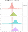
NPD (top) and penetration depth variability normalized by mean per device pooled across force levels (bottom). For NPD, the 0.0 y-axis value represents the skin surface; total n values/device are indicated in the top panel.
Figure 6.
Figure 6.
NPD (mean ± SD) versus skin application force per device; the 0.0 y-axis value represents the skin surface. Trend of deeper initial NPD at low forces and with increasing application forces observed for PN with posted-hub designs; n value 69 to 75 per device per force level. BD NanoTM 2nd Gen hub design more closely targets 4 mm PE length across the applied force range.
Figure 7.
Figure 7.
Depot depth density distribution for each device, pooled across force levels. Vertical lines indicate 4-4.5 mm depth range.
Figure 8.
Figure 8.
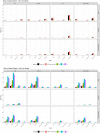
Observed erythema scores (0-4) for each device type graphed as a percentage of total injections pooled across applied force levels (left) and distributed within applied force levels (right).
Figure 9.
Figure 9.
Analysis of calculated ID and IM injection risk by test article using adult human skin thickness data categorized by gender, BMI, and injection site. The analysis includes a reference 4 mm ± 0.3 mm injection depth representing a physical needle length with no tissue compression factors (solid black bars).
Figure 10.
Figure 10.
Tissue compression following insertion of the NanoTM 2nd Gen PN (left) and posted-hub PN (right) into the tissue at a 1 lbf [4.4 N] application. In both images the orange line denotes PN hub/skin surface interface.

References

    1. Marcus A. Diabetes care—insulin delivery in a changing world. Medscape J Med. 2008;10:120. - PMC - PubMed
    1. Laurent A, Mistretta F, Bottigioli D, et al. Echographic measurement of skin thickness in adults by high frequency ultrasound to assess the appropriate microneedle length for intradermal delivery of vaccines. Vaccine. 2007;21;25:6423-6430. - PubMed
    1. Gibney MA, Arce CH, Byron KJ, Hirsch LJ. Skin and subcutaneous adipose layer thickness in adults with diabetes at sites used for insulin injections: implications for needle length recommendations. Curr Med Res Opin. 2010;26:1519-1530. - PubMed
    1. Ploin D, Schwarzenbach F, Dubray C, et al. Echographic measurement of skin thickness in sites suitable for intradermal vaccine injection in infants and children. Vaccine. 2011;29:8438-8442. - PubMed
    1. Ludescher B, Rommel M, Willmer T, Fritsche A, Schick F, Machann J. Subcutaneous adipose tissue thickness in adults—correlation with BMI and recommendations for pen needle lengths for subcutaneous self-injection. Clin Endocrinol (Oxf). 2011;75:786-790. - PubMed

Publication types