

microRNA 92b-3p regulates primordial follicle assembly by targeting TSC1 in neonatal mouse ovaries
- PMID: 30880550
- PMCID: PMC6527271
- DOI: 10.1080/15384101.2019.1593648
microRNA 92b-3p regulates primordial follicle assembly by targeting TSC1 in neonatal mouse ovaries
Abstract
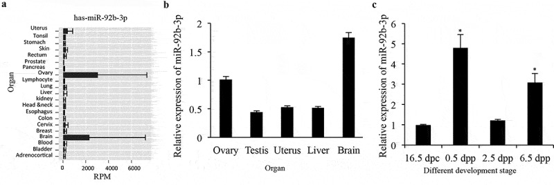
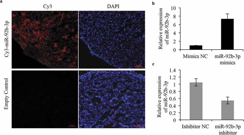
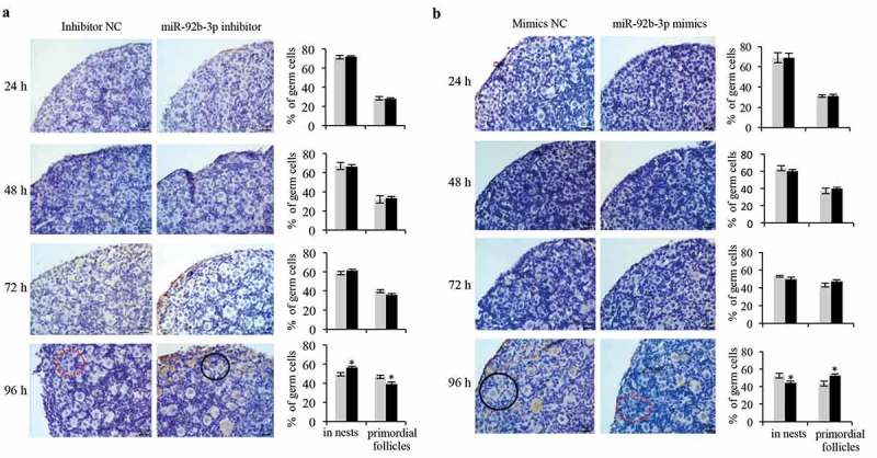
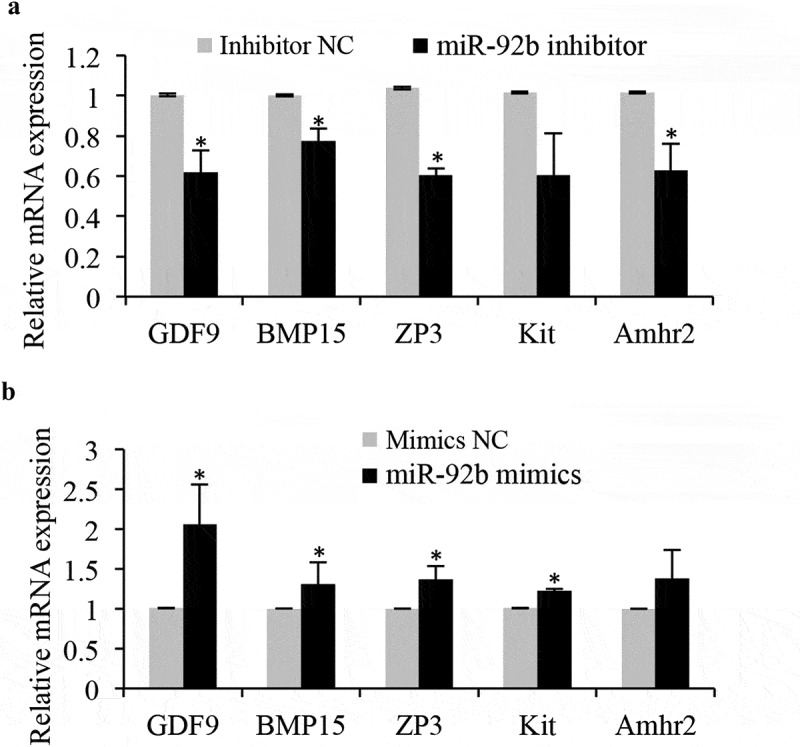
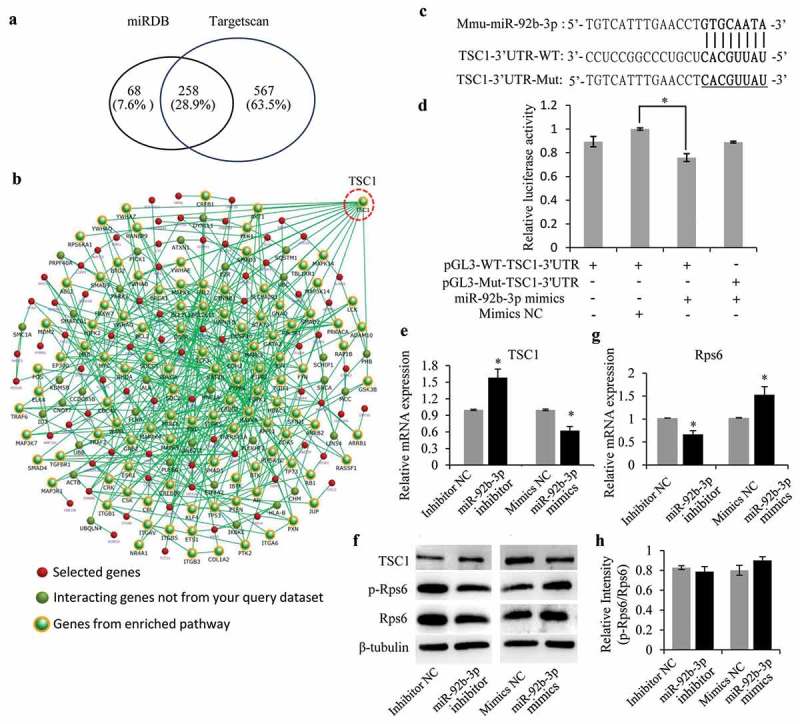
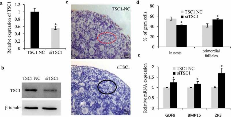
The primordial follicle pool, providing all oocytes available to a female throughout her reproductive life, is established perinatally. The formation of primordial follicle pool is regulated by precise transcriptional and post-transcriptional mechanisms. Recent studies have identified several microRNAs as post-transcriptional regulatory factors in the process of primordial follicle assembly. Here, we showed that miR-92b-3p was significantly upregulated in the stage of primordial follicle assembly in newborn mouse ovaries. Inhibiting miR-92b-3p suppressed the formation of primordial follicles, while overexpression of miR-92b-3p accelerated the processes of cyst breakdown and the following primordial follicle assembly. Accordingly, the expression of follicular development-related genes was reduced upon inhibiting of miR-92b-3p and increased under miR-92b-3p overexpression. Mechanistic studies identified TSC1 as a direct target of miR-92b-3p. miR-92b-3p could activate mTOR/Rps6 signaling through targeting and inhibiting TSC1 expression. In addition, knockdown of TSC1 showed an identical phenotype with that of miR-92b-3p overexpression in accelerating processes of cyst breakdown and primordial follicle formation. Thus, our work demonstrates that miR-92b-3p is a novel regulator of primordial follicle assembly by negatively regulating TSC1 in mTOR/Rps6 signaling.
Keywords: Mir-92b-3p; TSC1; mTOR; primordial follicle formation.
Figures
Similar articles
-
microRNA 376a regulates follicle assembly by targeting Pcna in fetal and neonatal mouse ovaries.Reproduction. 2014 Jul;148(1):43-54. doi: 10.1530/REP-13-0508. Epub 2014 Mar 31. Reproduction. 2014. PMID: 24686458
-
miR-92b-3p-TSC1 axis is critical for mTOR signaling-mediated vascular smooth muscle cell proliferation induced by hypoxia.Cell Death Differ. 2019 Sep;26(9):1782-1795. doi: 10.1038/s41418-018-0243-z. Epub 2018 Dec 5. Cell Death Differ. 2019. PMID: 30518907 Free PMC article.
-
MicroRNA-92b-3p is a prognostic oncomiR that targets TSC1 in clear cell renal cell carcinoma.Cancer Sci. 2020 Apr;111(4):1146-1155. doi: 10.1111/cas.14325. Epub 2020 Feb 19. Cancer Sci. 2020. PMID: 31975504 Free PMC article.
-
Cellular and molecular regulation of the activation of mammalian primordial follicles: somatic cells initiate follicle activation in adulthood.Hum Reprod Update. 2015 Nov-Dec;21(6):779-86. doi: 10.1093/humupd/dmv037. Epub 2015 Jul 30. Hum Reprod Update. 2015. PMID: 26231759 Review.
-
The impact of oocyte death on mouse primordial follicle formation and ovarian reserve.Reprod Med Biol. 2022 Nov 1;21(1):e12489. doi: 10.1002/rmb2.12489. eCollection 2022 Jan-Dec. Reprod Med Biol. 2022. PMID: 36329711 Free PMC article. Review.
Cited by
-
Ribosomal Protein S6: A Potential Therapeutic Target against Cancer?Int J Mol Sci. 2021 Dec 21;23(1):48. doi: 10.3390/ijms23010048. Int J Mol Sci. 2021. PMID: 35008473 Free PMC article. Review.
-
microRNAs Mediated Regulation of the Ribosomal Proteins and its Consequences on the Global Translation of Proteins.Cells. 2021 Jan 8;10(1):110. doi: 10.3390/cells10010110. Cells. 2021. PMID: 33435549 Free PMC article. Review.
-
Primordial Follicle Formation - Some Assembly Required.Curr Opin Endocr Metab Res. 2021 Jun;18:118-127. doi: 10.1016/j.coemr.2021.03.005. Epub 2021 Mar 20. Curr Opin Endocr Metab Res. 2021. PMID: 34027225 Free PMC article.
-
Extracellular vesicles derived from endometrial epithelial cells deliver exogenous miR-92b-3p to affect the function of embryonic trophoblast cells via targeting TSC1 and DKK3.Reprod Biol Endocrinol. 2022 Oct 25;20(1):152. doi: 10.1186/s12958-022-01023-z. Reprod Biol Endocrinol. 2022. PMID: 36284344 Free PMC article.
-
Long non-coding RNA Xist regulates oocyte loss via suppressing miR-23b-3p/miR-29a-3p maturation and upregulating STX17 in perinatal mouse ovaries.Cell Death Dis. 2021 May 25;12(6):540. doi: 10.1038/s41419-021-03831-4. Cell Death Dis. 2021. PMID: 34035229 Free PMC article.
References
-
- Pepling ME. From primordial germ cell to primordial follicle: mammalian femalegerm cell development. Genesis. 2006;44(12):622–632. - PubMed
-
- Pepling ME, Sundman EA, Patterson NL, et al. Differences in oocyte development and estradiol sensitivity among mouse strains. Reproduction. 2010;139(2):349–357. - PubMed
-
- Pepling ME, Spradling AC. Mouse ovarian germ cell cysts undergo programmed breakdown to form primordial follicles. Dev Biol. 2001;234(2):339–351. - PubMed
-
- Bristol-Gould SK, Kreeger PK, Selkirk CG, et al. Postnatal regulation of germ cells by activin: the establishment of the initial follicle pool. Dev Biol. 2006;298(1):132–148. - PubMed
-
- Zhang H, Adhikari D, Zheng W, et al. Combating ovarian aging depends on the use of existing ovarian follicles, not on putative oogonial stem cells. Reproduction. 2013;146(6):R229–233. - PubMed
Publication types
MeSH terms
Substances
LinkOut - more resources
Full Text Sources
Other Literature Sources
Miscellaneous