. 2019 Mar 1:14:525-534.
doi: 10.2147/CIA.S197444. eCollection 2019.

Aged kidneys are refractory to autophagy activation in a rat model of renal ischemia-reperfusion injury

Affiliations

Aged kidneys are refractory to autophagy activation in a rat model of renal ischemia-reperfusion injury

Changhui Diao et al. Clin Interv Aging. .

Abstract

Background: Ischemia-reperfusion (I/R) injury is the most common cause of acute kidney injury (AKI). Numerous therapeutic approaches for I/R injury have been studied, including autophagy, particularly in animal models of renal I/R injury derived from young or adult animals. However, the precise role of autophagy in renal ischemia-reperfusion in the aged animal model remains unclear. The purpose of this study was to demonstrate whether autophagy has similar effects on renal I/R injury in young and aged rats.

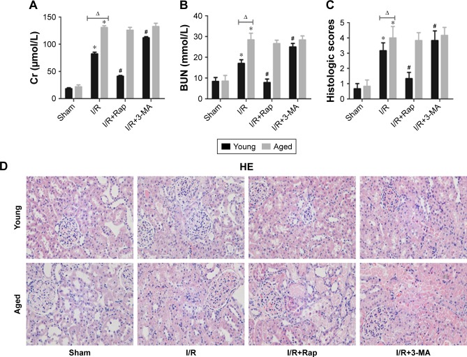
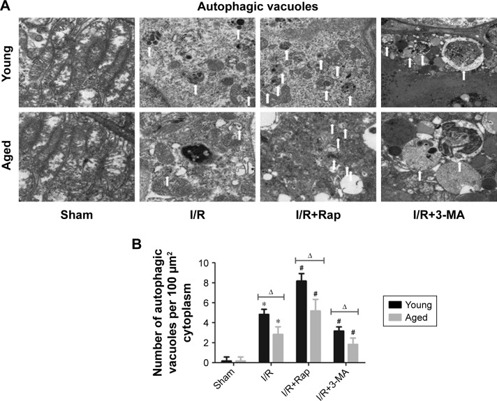
Materials and methods: All rats were divided into two age groups (3 months and 24 months) with each group being further divided into four subgroups (sham, I/R, I/R+Rap (rapamycin, an activator of autophagy), I/R+3-MA (3-methyladenine, an inhibitor of autophagy)). The I/R+Rap and I/R+3-MA groups were intraperitoneally injected with rapamycin and 3-MA prior to ischemia. We then measured serum levels of urea nitrogen, creatinine and assessed damage in the renal tissue. Immunohistochemistry was used to assess LC3-II and caspase-3, and Western blotting was used to evaluate the autophagy-related proteins LC3-II, Beclin-1 and P62. Apoptosis and autophagosomes were evaluated by TUNEL and transmission electron microscopy, respectively.

Results: Autophagy was activated in both young and aged rats by I/R and enhanced by rapamycin, although the level of autophagy was lower in the aged groups. In young rats, the activation of autophagy markedly improved renal function, reduced apoptosis in the renal tubular epithelial cells and the injury score in the renal tissue, thereby exerting protective effects on renal I/R injury. However, this level of protection was not present in aged rats.

Conclusion: Our data indicated that the activation of autophagy was ineffective in aged rat kidneys. These discoveries may have major implications in that severe apoptosis in aged kidneys might be refractory to antiapoptotic effect induced by the activation of autophagy.

Keywords: 3-methyladenine; aged; apoptosis; autophagy; ischemia-reperfusion; rapamycin; renal.


Conflict of interest statement

Disclosure The authors report no conflicts of interest in this work.

Figures

Figure 1
Figure 1
Effects of the enhancement and inhibition of autophagy upon renal function. Notes: SCr levels (A) and BUN levels (B). Scores for the histological appearance of acute tubular necrosis in young and aged rats (C). Representative images of rat kidney HE staining in young and aged rats (original magnification ×400) (D). The sham group showed normal renal cortical tissue structure. Obvious changes were observed in the I/R group and I/R+3-MA group, including acute tubular necrosis, tubule epithelial cell swelling, brush border loss, tubule dilation, cell shedding, tubule formation, and glomerular vascular congestion. Data are shown as the mean ± SD (n=6). *P<0.05 vs the same age sham-operated group; #P<0.05 vs same age I/R group; ΔP<0.05 vs young group under the same interventions. Abbreviations: 3-MA, 3-methyladenine; BUN, blood urea nitrogen; HE, hematoxylin-eosin; I/R, ischemia-reperfusion; Rap, rapamycin; SCr, serum creatinine.
Figure 2
Figure 2
Immunoblotting of LC3-II (A) (original magnification ×400). IOD/area indicating the expression of LC3-II (B). Both in young and aged groups, compared with I/R group, the IOD/area of LC3-II was higher when pretreated with rapamycin; the IOD/area of LC3-II was lower when pretreated with 3-MA. Data are shown as the mean ± SD (n=6). *P<0.05 vs same age sham-operated group; #P<0.05 vs same age I/R group; ΔP<0.05 vs young group under the same interventions. Abbreviations: 3-MA, 3-methyladenine; IOD, integrated optical density; I/R, ischemia-reperfusion; Rap, rapamycin.
Figure 3
Figure 3
Expressions of autophagy-related proteins in young and aged rats, including LC3-II/LC3-I, Beclin-1 and P62 (AC). Representative blots showing the effects of the enhancement and inhibition of autophagy upon active LC3-II/LC3-I, Beclin-1 and P62 expressions in the cytoplasm. The relative band densities of the three types of protein to the mean value of the control group in young and aged rats (DF). Data are shown as mean ± SD (n=6). *P<0.05 vs same age sham-operated group; #P<0.05 vs the same age I/R group; ΔP<0.05 vs young group under the same interventions. Abbreviations: 3-MA, 3-methyladenine; I/R, ischemia-reperfusion; Rap, rapamycin.
Figure 4
Figure 4
High magnification electron micrographs showing ultrastructure changes (A) (sham, I/R, I/R+Rap, and I/R+3-MA groups, original magnification ×1,700; I/R+3-MA groups, original magnification ×5,000). Autophagic vacuoles (white arrow) containing whorls of membranous material and some cytoplasm. In the young and aged groups, the most numerous double-membrane autophagic vacuoles were found in the I/R+Rap group, flowed by the I/R group; the least number of vacuoles was found in the sham group. Quantification of the number of autophagic vacuoles per 100 µm2 cytoplasm (B). Notes: Data are shown as mean ± SD (n=6). *P<0.05 vs the same age sham-operated group; #P<0.05 vs same age I/R group; ΔP<0.05 vs young group under the same interventions. Abbreviations: 3-MA, 3-methyladenine; I/R, ischemia-reperfusion; Rap, rapamycin.
Figure 5
Figure 5
Immunofluorescent image of TUNEL staining for apoptotic cells in kidney tissue (A) (original magnification ×400) and immunoblotting of caspase-3 (B) (original magnification ×400). Nuclei of TUNEL-positive cells are stained red. Mean number of TUNEL-positive cells per high power field (HPF ×400) (C). IOD/area indicated the expression of caspase-3 (D). Data are shown as mean ± SD (n=6). *P<0.05 vs the same age sham-operated group; #P<0.05 vs same age I/R group; ΔP<0.05 vs young group under the same interventions. Abbreviations: 3-MA, 3-methyladenine; IOD, integrated optical density; I/R, ischemia-reperfusion; Rap, rapamycin.

Similar articles

Cited by

References

    1. Matsumoto T, Urushido M, Ide H, et al. Small heat shock protein Beta-1 (HSPB1) is upregulated and regulates autophagy and apoptosis of renal tubular cells in acute kidney injury. PLoS One. 2015;10(5):e0126229. - PMC - PubMed
    1. Mehta RL, Burdmann EA, Cerdá J, et al. Recognition and management of acute kidney injury in the International Society of nephrology 0 by 25 global snapshot: a multinational cross-sectional study. Lancet. 2016;387(10032):2017–2025. - PubMed
    1. Kunzendorf U, Haase M, Rölver L, Haase-Fielitz A. Novel aspects of pharmacological therapies for acute renal failure. Drugs. 2010;70(9):1099–1114. - PubMed
    1. Venkatachalam MA, Weinberg JM, Kriz W, Bidani AK. Failed tubule recovery, AKI-CKD transition, and kidney disease progression. J Am Soc Nephrol. 2015;26(8):1765–1776. - PMC - PubMed
    1. Xie Y, Xiao J, Fu C, Zhang Z, Ye Z, Zhang X. Ischemic preconditioning promotes autophagy and alleviates renal ischemia/reperfusion injury. Biomed Res Int. 2018;2018:1–11. - PMC - PubMed

MeSH terms