

Iron in Neurodegeneration - Cause or Consequence?
- PMID: 30881284
- PMCID: PMC6405645
- DOI: 10.3389/fnins.2019.00180
Iron in Neurodegeneration - Cause or Consequence?
Abstract
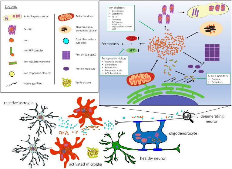
Iron dyshomeostasis can cause neuronal damage to iron-sensitive brain regions. Neurodegeneration with brain iron accumulation reflects a group of disorders caused by iron overload in the basal ganglia. High iron levels and iron related pathogenic triggers have also been implicated in sporadic neurodegenerative diseases including Alzheimer's disease (AD), Parkinson's disease (PD), and multiple system atrophy (MSA). Iron-induced dyshomeostasis within vulnerable brain regions is still insufficiently understood. Here, we summarize the modes of action by which iron might act as primary or secondary disease trigger in neurodegenerative disorders. In addition, available treatment options targeting brain iron dysregulation and the use of iron as biomarker in prodromal stages are critically discussed to address the question of cause or consequence.
Keywords: autophagic-lysosomal dysfunction; iron; mitochondrial dysfunction; neurodegeneration; neurodegenerative mechanisms; neuroinflammation; protein aggregation.
Figures
References
-
- Ahmadi S., Ebralidze I. I., She Z., Kraatz H.-B. (2017). Electrochemical studies of tau protein-iron interactions—Potential implications for Alzheimer’s Disease. Electrochim. Acta 236 384–393. 10.1016/j.electacta.2017.03.175 - DOI
Publication types
LinkOut - more resources
Full Text Sources