. 2013 Dec 24;3(4):e24.
doi: 10.2106/JBJS.ST.M.00041. eCollection 2014 Dec.

Limb Lengthening with a Submuscular Locking Plate

Affiliations

Limb Lengthening with a Submuscular Locking Plate

Chang-Wug Oh et al. JBJS Essent Surg Tech. .

Abstract

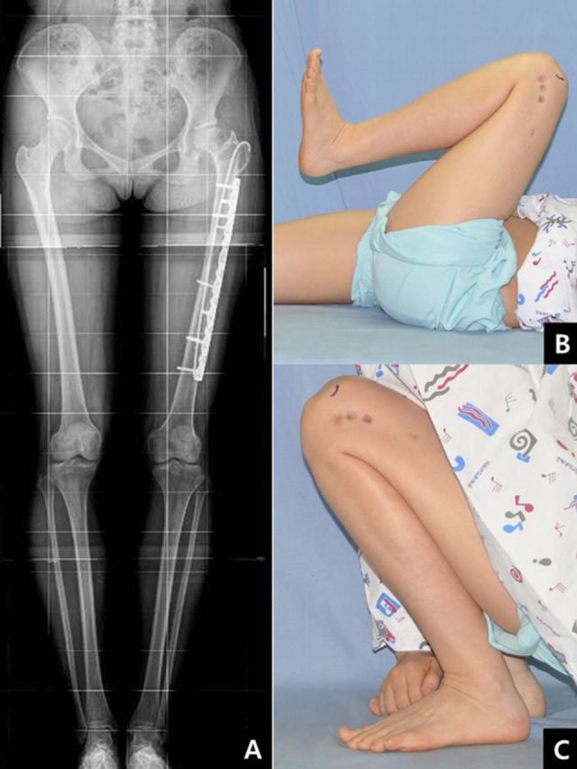
Introduction: Limb-lengthening with a submuscular locking plate provides a good alternative for patients, especially children, in whom lengthening over an intramedullary nail would be difficult.

Step 1 external fixation for lengthening with submuscular plating: The first operation is divided into two steps, submuscular plating and external fixation with corticotomy; due to the anatomical characteristics, the procedures differ in the tibia and femur.

Step 2 lengthening: Start distraction at seven to ten days after surgery and continue until the target length is achieved.

Step 3 locking of the distal segment and removal of the external fixator: When the target length has been achieved, place screws into the distal segment through plate holes and remove the fixator.

Step 4 postoperative care: Start with partial weight-bearing, obtain a radiograph every four to eight weeks, and allow full weight-bearing with crutches when osseous consolidation is observed.

Results: We prospectively performed limb lengthening using an external fixator and a submuscular locking plate in ten patients16.IndicationsContraindicationsPitfalls & Challenges.


Figures

Fig. 1
Fig. 1
An eleven-year old boy had an idiopathic leg-length discrepancy due to tibial shortening of about 32 mm. (Reproduced, with permission and copyright of British Society of Bone and Joint Surgery, from: Oh CW, Song HR, Kim JW, Choi JW, Min WK, Park BC. Limb lengthening with a submuscular locking plate. J Bone Joint Surg Br. 2009 Oct;91[10]:1394-9.)
Fig. 2
Fig. 2
After fibular osteotomy, a submuscular locking plate was placed laterally, and four locking screws were fixed to the proximal segment (Figs. 2-A, 2-B, and 2-C). Then, a monolateral external fixator for lengthening was placed medially and percutaneous corticotomy was performed (Fig. 2-D).
Fig. 3
Fig. 3
Schematic illustration of the postoperative status (Fig. 3-A), a postoperative radiograph (Fig. 3-B), and a clinical photograph (Fig. 3-C). Note that the locking screws and Schanz pin should not intrude upon the physeal plate. (Figs. 3-B and 3-C reproduced, with permission and copyright of British Society of Bone and Joint Surgery, from: Oh CW, Song HR, Kim JW, Choi JW, Min WK, Park BC. Limb lengthening with a submuscular locking plate. J Bone Joint Surg Br. 2009 Oct;91[10]:1394-9.)
Fig. 4
Fig. 4
Distraction was performed until the target length was achieved. (Reproduced, with permission and copyright of British Society of Bone and Joint Surgery, from: Oh CW, Song HR, Kim JW, Choi JW, Min WK, Park BC. Limb lengthening with a submuscular locking plate. J Bone Joint Surg Br. 2009 Oct;91[10]:1394-9.)
Fig. 5
Fig. 5
After lengthening was done, locking of the distal segment was performed in a percutaneous manner, after which the external fixator was removed.
Fig. 6
Fig. 6
Postoperative radiograph (Fig. 6-A) and photograph showing the wound (Fig. 6-B). (Reproduced, with permission and copyright of British Society of Bone and Joint Surgery, from: Oh CW, Song HR, Kim JW, Choi JW, Min WK, Park BC. Limb lengthening with a submuscular locking plate. J Bone Joint Surg Br. 2009 Oct;91[10]:1394-9.)
Fig. 7
Fig. 7
Six months later, the distraction callus had healed (Figs. 7-A and 7-B) and the patient had an excellent functional result (Fig. 7-C). (Figs. 7-A and 7-B reproduced, with permission and copyright of British Society of Bone and Joint Surgery, from: Oh CW, Song HR, Kim JW, Choi JW, Min WK, Park BC. Limb lengthening with a submuscular locking plate. J Bone Joint Surg Br. 2009 Oct;91[10]:1394-9.)
Fig. 8
Fig. 8
A twenty-nine-year-old woman had femoral shortening of 42 mm as a result of septic hip sequelae.
Fig. 9
Fig. 9
A submuscular locking plate was fixed with a flexible intramedullary nail after corticotomy. Note the three half-pins of the external fixator fixed proximal and distal to the plate.
Fig. 10
Fig. 10
After achievement of the target length (Fig. 10-A), screws were fixed percutaneously at the distal segment (Fig. 10-B). The external fixator was removed at this stage.
Fig. 11
Fig. 11
Postoperative radiographs (Figs. 11-A and 11-B) and a photograph showing the wound (Fig. 11-C).
Fig. 12
Fig. 12
One year later, the distraction callus had healed.
Fig. 13
Fig. 13
The lengths of both lower limbs were the same (Fig. 13-A), and the functional result was excellent (Figs. 13-B and 13-C).

References

    1. Ilizarov GA. The principles of the Ilizarov method. Bull Hosp Jt Dis Orthop Inst. 1988 Spring;48(1):1-11. - PubMed
    1. Price CT Mann JW. Experience with the Orthofix device for limb lengthening. Orthop Clin North Am. 1991 Oct;22(4):651-61. - PubMed
    1. Stanitski DF Shahcheraghi H Nicker DA Armstrong PF. Results of tibial lengthening with the Ilizarov technique. J Pediatr Orthop. 1996 Mar-Apr;16(2):168-72. - PubMed
    1. Zarzycki D Tesiorowski M Zarzycka M Kacki W Jasiewicz B. Long-term results of lower limb lengthening by the Wagner method. J Pediatr Orthop. 2002 May-Jun;22(3):371-4. - PubMed
    1. Paley D. Problems, obstacles, and complications of limb lengthening by the Ilizarov technique. Clin Orthop Relat Res. 1990 Jan;(250):81-104. - PubMed